. 2014 Jul;44(7):986-98.
doi: 10.1111/cea.12324.

Grass-specific CD4(+) T-cells exhibit varying degrees of cross-reactivity, implications for allergen-specific immunotherapy

Affiliations

Grass-specific CD4(+) T-cells exhibit varying degrees of cross-reactivity, implications for allergen-specific immunotherapy

L D Archila et al. Clin Exp Allergy. 2014 Jul.

Abstract

Background: Conceptually, allergic responses may involve cross-reactivity by antibodies or T-cells. While IgE cross-reactivity among grass-pollen allergens has been observed, cross-reactivity at the allergen-specific T-cell level has been less documented. Identification of the patterns of cross-reactivity may improve our understanding, allowing optimization of better immunotherapy strategies.

Objectives: We use Phleum pratense as model for the studying of cross-reactivity at the allergen-specific CD4(+) T cell level among DR04:01 restricted Pooideae grass-pollen T-cell epitopes.

Methods: After in vitro culture of blood mono-nucleated cells from grass-pollen-allergic subjects with specific Pooideae antigenic epitopes, dual tetramer staining with APC-labelled DR04:01/Phleum pratense tetramers and PE-labelled DR04:01/Pooideae grass homolog tetramers was assessed to identify cross-reactivity among allergen-specific DR04:01-restricted T-cells in six subjects. Direct ex vivo staining enabled the comparison of frequency and phenotype of different Pooideae grass-pollen reactive T-cells. Intracellular cytokine staining (ICS) assays were also used to examine phenotypes of these T-cells.

Results: T-cells with various degrees of cross-reactive profiles could be detected. Poa p 1 97-116 , Lol p 1 221-240 , Lol p 5a 199-218 , and Poa p 5a 199-218 were identified as minimally cross-reactive T-cell epitopes that do not show cross-reactivity to Phl p 1 and Phl p 5a epitopes. Ex vivo tetramer staining assays demonstrated T-cells that recognized these minimally cross-reactive T-cell epitopes are present in Grass-pollen-allergic subjects.

Conclusions: Our results suggest that not all Pooideae grass epitopes with sequence homology are cross-reactive. Non-cross-reactive T-cells with comparable frequency, phenotype and functionality to Phl p-specific T-cells suggest that a multiple allergen system should be considered for immunotherapy instead of a mono-allergen system.

Keywords: CD4+; MHC class II tetramers; Pooideae; T-cells; allergy; cross-reactivity; epitopes; grass-pollen.


Figures

Figure 1
Figure 1
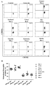
Subjects allergic to TGP have IgE sensitivity to different grass-species. Basophils were stimulated with pollen extracts from 6 different Pooideae species or a mix containing grass-pollen from all species (Greer Laboratories, USA) and were stained simultaneously with a panel of antibodies of interest. Upregulation of CD203c indicated activation of basophils. A. Representative result for Allergy #89. B. Percentages of CD203c on stimulated basophils are summarized for each DR04:01 allergic subject.
Figure 2
Figure 2
Grass-specific CD4+ T-cells exhibit varying degrees of cross-reactivity. After in vitro stimulation with Phl p homologous grass peptides (top panels of A, B and C, amino-acid sequence of the stimulating peptide is specified on top of each panel) or Phl p 167-186 AAFKVAATAANAA, bottom panels of A, B and C), cells were co-stained with APC-labeled DR04:01/Phl p grass peptide-loaded tetramers (Y axis) and with PE-labeled DR04:01/homologous grass peptide-loaded tetramers corresponding to the stimulating epitopes (X axis). The plots in A, B and C show representative results for different cross-reactivity profiles for the TGP-derived epitopes in the Group 5a 167-186 region. A. Full-scale cross-reactivity was observed for Poa p 5a 167-186, and Pha a 5a 167-186. B. Partial cross-reactivity was observed for Dac g 5a 167-186 and Hol 5a 167-186. C. Minimal cross-reactivity was observed for Lol p 5a 167-186 and Hor v 5a 167-186. D. Comparison of percentages of cross-reactive T-cells for Group 1 homologs. E. Comparison of percentages of cross-reactive T-cells for Group 5a homologs. Summarized results from (n=6) allergic subjects are presented for distinct antigenic epitope regions. Each bar depicts the average of dual-tetramer stained T-cells (cross-reactive) observed per epitope.
Figure 3
Figure 3
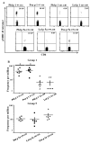
Minimally cross-reactive CD4+ T-cells can be detected ex vivo in TGP allergic subjects. Determination of frequencies of Phl p- and grass homolog-reacting CD4+ T-cells for different regions of Group 1 and 5a allergens. A. Example of ex-vivo tetramer staining for TGP–derived Group 1 97-116, Group 1 221-240 and Group 5a 97-116-Phl p-specific and homologous minimally cross-reactive CD4+ T-cells from a grass pollen allergic subject with an ImmunoCAP score of 5 for Phl p specific-IgE. B. Frequencies for TGP-derived Group 1 97-116 and Group 1 221-240, and Group 5a 199-218 grass homolog-reactive CD4+ T-cells. Each data point denotes the frequency of epitope specific CD4+ T-cells for a different individual. A t student test was used in the statistical analysis.*P < 0.05 to compare populations from different epitopes.
Figure 4
Figure 4
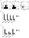
Minimally cross-reactive CD4+ T-cells are memory T-cells with a TH2 phenotype and are activated during the pollen-season. A. Anti-PE enrichment permitted the analysis of surface marker expression; a naïve marker (CD45RA), a TH2 marker (CCR4) and an activation marker (CD38) were analyzed to compare Phl p grass-specific CD4+ T-cells with homologous grass-specific CD4+ T-cells. PBMC from a TPG allergic subject were stained with Poa p corresponding for TGP–derived region Group 1 97-116 and are presented as an example. B. Each bar represents surface expression of different markers for Group 1 reactive T-cells for 6 different allergic subjects. C. Each bar represents surface expression of different markers for Group 5 reactive T-cells for 6 different subjects.
Figure 5
Figure 5
Minimally cross-reactive CD4+ T-cells functional profiles correlate with a TH2 dominated response. An example of intracellular cytokine staining for Phl p- and grass homolog-reactive T-cells is provided. A. Representative result for ICS. PBMC from a TGP allergic subject were stimulated with Poa p peptide corresponding to region Group 1 97-116 and cultured for 2 weeks, cells were than stained with the corresponding PE-labeled DR04:01/Poa p grass peptide-loaded tetramers. Tetramer+ cells were gated and dual cytokine analysis is presented. B. Summarized results of ICS for TGP-derived Group 1 97-116, Group 1 221-240 and Group 5 199-218 epitopes, each bar depicts percentage of tetramer-positive cytokine producing cells in each individual.
Figure 6
Figure 6
Grass-pollen-specific CD4+ T-cells have varying degrees of cross-reactivity that vary on the HLA and epitope they recognize. A. PBMC (n=2) from DR07:01 allergic subjects were stimulated in vitro with Dac g peptide corresponding to region Group 5b 89-108. Cells were then co-stained with APC-labeled DR07:01/Phl p grass peptide-loaded tetramers (Y axis) as well as PE-labeled DR0701/Dac g 5b peptide-loaded tetramers (X axis). The plot shows a representative result for Group 5 89-108 grass homologs, sequence of stimulating peptide is shown on top of plot. T-cells were gated on Sidescatter and CD4+. The graph represents percentage of T-cells that were able to double stain; for this particular HLA-restriction minimal cross-reactivity was detected stimulating with either Dac g 5b or Phl p 5b peptides (about 20% of T-cells double stained) B. Ex vivo frequency of allergen-specific CD4+ T-cells for Group 5b 89-108 homologs. C. PBMC (n=2) from DRB5 allergic subjects were stimulated in vitro with either Dac g peptide corresponding to region Group 5b 152-160. Cells where then co-stained with APC-labeled DRB5/Phl p grass peptide-loaded tetramers (Y axis) as well as PE-labeled DRB5/Dac g 5b peptide-loaded tetramers (X axis). The plot shows a representative result for Group 5b 152-160 grass homologs, sequence of stimulating peptide is shown on top of plot. T-cells were gated on Side scatter and CD4+. The graph represents percentage of double stained T-cells, for this particular HLA-restriction full scale cross-reactivity was detected stimulating with either Dac g 5b or Phl p 5b peptides (≥ 90% of T-cells double stained) D. Ex vivo frequency of allergen-specific CD4+ T-cells for Group 5 152-160 grass homologs.

Comment in

Similar articles

Cited by

References

    1. Weber RW. Patterns of pollen cross-allergenicity. J Allergy Clin Immunol. 2003;112:229–39. - PubMed
    1. Weber RW. Cross-reactivity of pollen allergens. Curr Allergy Asthma Rep. 2004;4:401–8. - PubMed
    1. White JF, Bernstein DI. Key pollen allergens in North America. Ann Allergy Asthma Immunol. 2003;91:425–35. - PubMed
    1. Weber RW, Nelson HS. Pollen allergens and their interrelationships. Clin Rev Allergy. 1985;3:291–318. - PubMed
    1. Marcucci F, Sensi L, Di CG, Incorvaia C, Puccinelli P, Scurati S, Frati F. Which allergen extract for grass pollen immunotherapy? An in vitro study. Immunol Invest. 2010;39:635–44. - PubMed

Publication types

MeSH terms