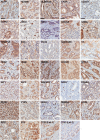
Evaluation of protein biomarkers of prostate cancer aggressiveness
- PMID: 24708576
- PMCID: PMC4101830
- DOI: 10.1186/1471-2407-14-244
Evaluation of protein biomarkers of prostate cancer aggressiveness
Abstract
Background: Prognostic multibiomarker signatures in prostate cancer (PCa) may improve patient management and provide a bridge for developing novel therapeutics and imaging methods. Our objective was to evaluate the association between expression of 33 candidate protein biomarkers and time to biochemical failure (BF) after prostatectomy.
Methods: PCa tissue microarrays were constructed representing 160 patients for whom clinicopathologic features and follow-up data after surgery were available. Immunohistochemistry for each of 33 proteins was quantified using automated digital pathology techniques. Relationships between clinicopathologic features, staining intensity, and time to BF were assessed. Predictive modeling using multiple imputed datasets was performed to identify the top biomarker candidates.
Results: In univariate analyses, lymph node positivity, surgical margin positivity, non-localized tumor, age at prostatectomy, and biomarkers CCND1, HMMR, IGF1, MKI67, SIAH2, and SMAD4 in malignant epithelium were significantly associated with time to BF. HMMR, IGF1, and SMAD4 remained significantly associated with BF after adjusting for clinicopathologic features while additional associations were observed for HOXC6 and MAP4K4 following adjustment. In multibiomarker predictive models, 3 proteins including HMMR, SIAH2, and SMAD4 were consistently represented among the top 2, 3, 4, and 5 most predictive biomarkers, and a signature comprised of these proteins best predicted BF at 3 and 5 years.
Conclusions: This study provides rationale for investigation of HMMR, HOXC6, IGF1, MAP4K4, SIAH2, and SMAD4 as biomarkers of PCa aggressiveness in larger cohorts.
Figures


References
-
- Cookson MS, Aus G, Burnett AL, Canby-Hagino ED, D’Amico AV, Dmochowski RR, Eton DT, Forman JD, Goldenberg SL, Hernandez J, Higano CS, Kraus SR, Moul JW, Tangen C, Thrasher JB, Thompson I. Variation in the definition of biochemical recurrence in patients treated for localized prostate cancer: the American Urological Association Prostate Guidelines for Localized Prostate Cancer Update Panel report and recommendations for a standard in the reporting of surgical outcomes. J Urol. 2007;177(2):540–545. doi: 10.1016/j.juro.2006.10.097. - DOI - PubMed
-
- Frazier HA, Robertson JE, Humphrey PA, Paulson DF. Is prostate specific antigen of clinical importance in evaluating outcome after radical prostatectomy. J Urol. 1993;149(3):516–518. - PubMed
Publication types
MeSH terms
Substances
Grants and funding
LinkOut - more resources
Full Text Sources
Other Literature Sources
Medical
Research Materials
Miscellaneous

