. 2014 Apr 5:11:71.
doi: 10.1186/1742-2094-11-71.

Epidermal growth factor receptor inhibitor ameliorates excessive astrogliosis and improves the regeneration microenvironment and functional recovery in adult rats following spinal cord injury

Affiliations

Epidermal growth factor receptor inhibitor ameliorates excessive astrogliosis and improves the regeneration microenvironment and functional recovery in adult rats following spinal cord injury

Zai-Wang Li et al. J Neuroinflammation. .

Abstract

Background: Astrogliosis is a common phenomenon after spinal cord injury (SCI). Although this process exerts positive effects on axonal regeneration, excessive astrogliosis imparts negative effects on neuronal repair and recovery. Epidermal growth factor receptor (EGFR) pathway is critical to the regulation of reactive astrogliosis, and therefore is a potential target of therapeutics to better control the response. In this report, we aim to investigate whether blocking EGFR signaling using an EGFR tyrosine kinase specific inhibitor can attenuate reactive astrogliosis and promote functional recovery after a traumatic SCI.

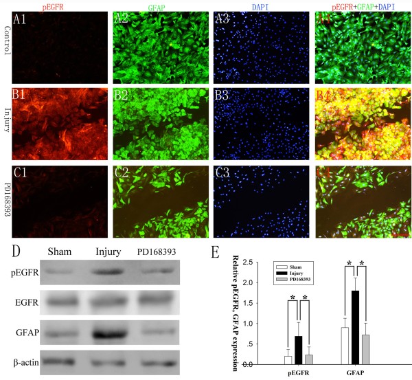
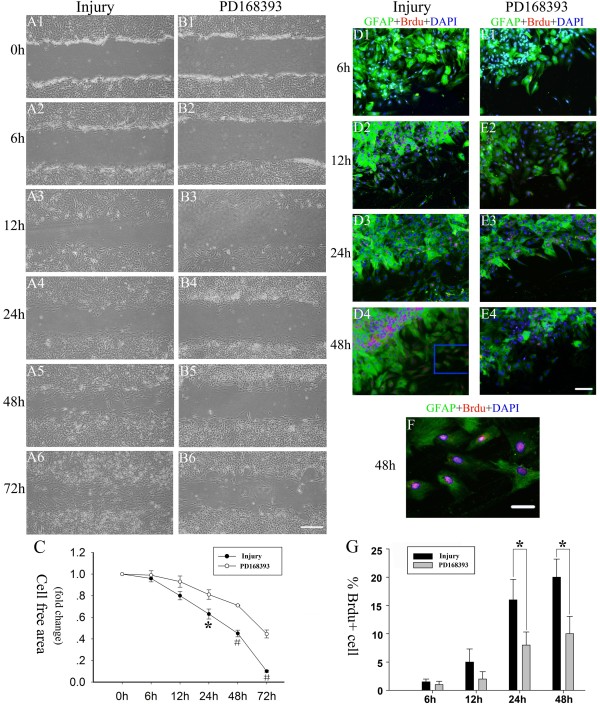
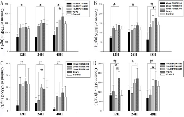
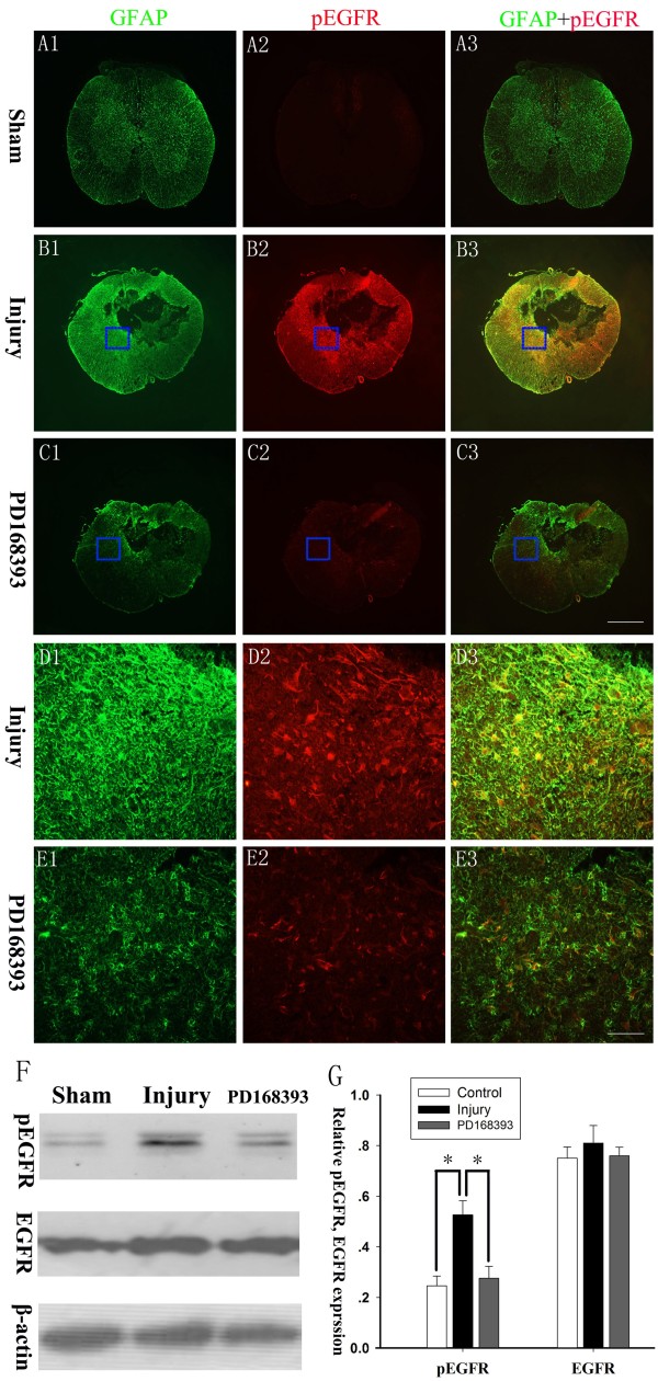
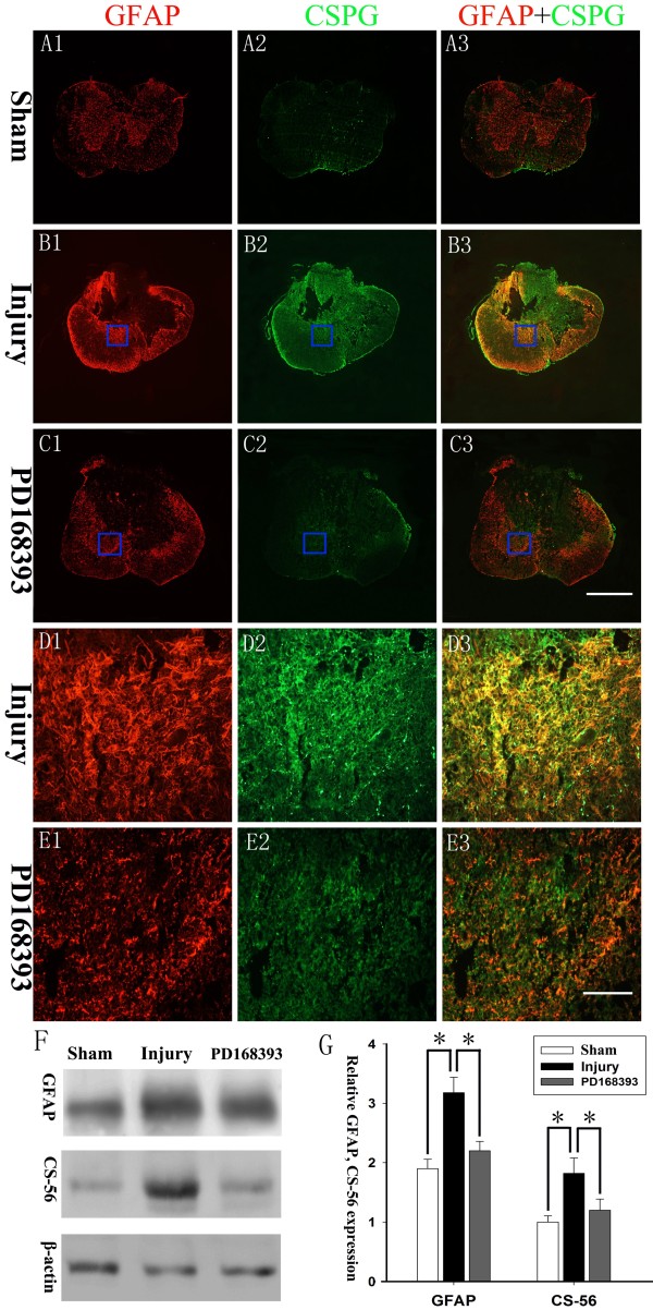
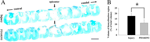
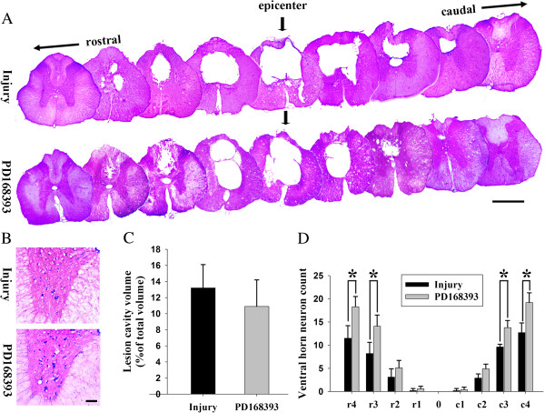
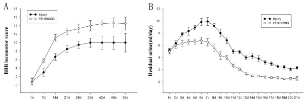
Method: The astrocyte scratch injury model in vitro and the weight-drop SCI model in vivo were used as model systems. PD168393 was used to inhibit EGFR signaling activation. Astrocytic activation and phosphorylated EGFR (pEGFR) were observed after immunofluorescence staining and Western blot analysis. The rate of proliferation was determined by immunofluorescence detection of BrdU-incorporating cells located next to the wound. The levels of TNF-α, iNOS, COX-2 and IL-1β in the culture medium under different conditions were assayed by ELISA. Western blot was performed to semi-quantify the expression of EGFR/pEGFR, glial fibrillary acid protein (GFAP) and chondroitin sulfate proteoglycans (CSPGs). Myelin was stained by Luxol Fast Blue Staining. Cresyl violet eosin staining was performed to analyze the lesion cavity volume and neuronal survival following injury. Finally, functional scoring and residual urine recording were performed to show the rats' recovery.

Results: EGFR phosphorylation was found to parallel astrocyte activation, and EGFR inhibitor PD168393 potently inhibited scratch-induced reactive astrogliosis and proinflammatory cytokine/mediator secretion of reactive astrocytes in vitro. Moreover, local administration of PD168393 in the injured area suppressed CSPGs production and glial scar formation, and resulted in reduced demyelination and neuronal loss, which correlated with remarkable hindlimb motor function and bladder improvement in SCI rats.

Conclusions: The specific EGFR inhibitor PD168393 can ameliorate excessive reactive astrogliosis and facilitate a more favorable environment for axonal regeneration after SCI. As such, EGFR inhibitor may be a promising therapeutic intervention in CNS injury.


Figures

Figure 1
Figure 1
PD168393 inhibited EGFR phosphorylation of astrocytes in an in vitro scratch wound model. Cultured astrocytes were immunostained for pEGFR, GFAP and DAPI in control (A1-A4), injury, (B1-B4) and PD168393 (40 μM) treatment groups (C1-C4) at 24 hours after injury (n = 5 in each group). (A1-C1) immunolabeling for pEFGR, (A2-C2), GFAP, (A3-C3) DAPI and (A4-C4) for co-localization of pEGFR, GFAP and DAPI. Scale bars = 100 μm. Representative Western blots of pEGFR and GFAP expression are shown (n = 3/group) (D). Semiquantitative analysis of pEGFR as a ratio of EGFR loading and semiquantitative measurements of GFAP were obtained by normalization to β-actin (E). *P < 0.05.
Figure 2
Figure 2
PD168393 treatment slowed down scratch injury-induced reactive astrogliosis. Astrocytes were scratched and incubated in the absence or with treatment of 40 μM PD168393. (A1-B1), (A2-B2), (A3-B3), (A4-B4), (A5-B5) and (A6-B6) are paired images taken from control 0, 6, 12, 24, 48 and 72 hour groups after scratch injury, respectively. Scale bars = 100 μm in B6 (applies to A1-B6). Graphical representation of remaining cell-free area quantification over 72 hours after scratch injury (C). Data are expressed as fold change compared to control group (0 hours). Values are expressed means ± SD (n = 5). Significant difference between time points was observed (*P < 0.05, #P < 0.01). Immunostaining for Brdu, glial fibrillary acid protein (GFAP) and 4,6-diamidino-2-phenylindole (DAPI) at 6, 12, 24 and 48 hours after scratch injury in the injury group (D1-D4) and PD168393 (40 μM) treatment group (E1-E4), respectively. Astrocytes were identified by GFAP immunostaining (green) and nuclei were identified by DAPI labeling (blue). Astrocytes in S phase were identified by BrdU labeling (red). Insets in panels (D4) of co-localization of phosphorylated epidermal growth factor receptor (pEGFR), GFAP and DAPI are shown at high magnification in (F). Scale bars = 100 μm in E4 (applies to D1-E4); scale bars = 20 μm in (F). Percentage of astroglial cells with BrdU labeling at different times after scratch injury (G). *P < 0.05.
Figure 3
Figure 3
PD168393 inhibited proinflammatory cytokine/mediators secretion of astrocytes in a scratch wound model. TNF-α (A), iNOS (B), COX-2 (C) and IL-1β (D) in conditioned medium from control, injury and PD168393 groups (at 10 μM, 20 μM and 40 μM concentration) were assayed by ELISA at various time intervals (12, 24 and 48 hours after injury). *P < 0.05, #P < 0.01.
Figure 4
Figure 4
PD168393 inhibited epidermal growth factor receptor (EGFR) phosphorylation of astrocytes after spinal cord injury (SCI) in rats. Spinal cord sections were immunostained for phosphorylated EGFR (pEGFR) and glial fibrillary acid protein (GFAP) at two weeks after SCI (n = 5/group). (A1-C1) GFAP, (A2-C2) pEFGR, and (A3-C3) co-localization of pEGFR and GFAP. Insets in panels (B1-C3) are shown at high magnification in panels (D1-E3), respectively. Scale bars = 500 μm in (C3) (applies to A1-C3); scale bars = 50 μm in (E3) (applies to D1-E3). Representative Western blots of pEGFR and EGFR expression (n = 3/group), and β-actin was a loading control (F). Semi-quantitative measurements were obtained by normalizing to β-actin (G). *P < 0.05.
Figure 5
Figure 5
PD168393 attenuated chondroitin sulfate proteoglycan (CSPG) production from reactive astrocytes after spinal cord injury (SCI). Double-immunofluorescent labeling for CSPGs and glial fibrillary acid protein (GFAP) at four weeks after SCI (n = 5/group). (A1-C1) GFAP, (A2-C2) CSPG, and (A3-C3) co-localization of CSPG and GFAP. Insets in panels (B1-C3) are shown at high magnification in panels (D1-E3). Scale bars = 500 μm in (C3) (applies to A1-C3); scale bars = 50 μm in (E3) (applies to D1-E3). Representative Western blots of GFAP and CS-56 expression (n = 3/group), and β-actin was a loading control (F). Semi-quantitative measurements were obtained by normalizing β-actin loading (G). *P < 0.05.
Figure 6
Figure 6
PD168393 reduced demyelination after spinal cord injury (SCI). A 15 mm length of spinal cord centered at the injury site was harvested at day 28 post-injury and serially sectioned (n = 5/group). Representative transverse Luxol Fast Blue stained sections at the epicenter and in 1 mm increments rostral and caudal to the epicenter. Epicenter sections are indicated by arrows. Scale bar = 1 mm (A). Graphical representation shows statistically significant reduction in lesion cavity volume following PD168393 treatment as well as considerable decrease in myelin loss (B) (n = 5, *P < 0.05 compared to control).
Figure 7
Figure 7
PD168393 promoted survival of ventral horn (VH) motor neurons after spinal cord injury (SCI). Representative transverse cresyl violet eosin stained sections of four weeks post-SCI at the epicenter and in 1 mm increments rostral and caudal to the epicenter. Epicenter sections are indicated by arrows. Scale bar = 1 mm (A). Representative photomicrographs from four weeks post-SCI showing cresyl violet eosin stained VH neurons at 4 mm rostral to the injury epicenter. Scale bar = 50 μm (B). Graphical representation showing no statistically significant difference in lesion cavity volume following treatment (n = 5) (C). Comparison of VH neurons among different groups at various distances from the injury epicenter (0) as well as 1 to 4 mm rostral (r) and caudal (c) to it (D) (n = 5, *P < 0.05).
Figure 8
Figure 8
PD168393 improved functional outcome and bladder recovery after spinal cord injury (SCI). The Basso, Bresnahan, and Beattie (BBB) scores of hindlimb locomotion were assessed at day 1 post-injury and weekly thereafter. Repeated-measures ANOVA revealed significant difference between injury and PD168393-treated animals (P < 0.01) (A). The PD168393-treated rats had less residual urine compared with non-treated injured rats. Statistical analysis between animal groups showed significant differences (P < 0.05) (B) (n = 5/group).

Similar articles

Cited by

References

    1. Grimpe B, Silver J. A novel DNA enzyme reduces glycosaminoglycan chains in the glial scar and allows microtransplanted dorsal root ganglia axons to regenerate beyond lesions in the spinal cord. J Neurosci. 2004;24:1393–1397. doi: 10.1523/JNEUROSCI.4986-03.2004. - DOI - PMC - PubMed
    1. Davies JE, Tang X, Denning JW, Archibald SJ, Davies SJ. Decorin suppresses neurocan, brevican, phosphacan and NG2 expression and promotes axon growth across adult rat spinal cord injuries. Eur J Neurosci. 2004;19:1226–1242. doi: 10.1111/j.1460-9568.2004.03184.x. - DOI - PubMed
    1. Usher LC, Johnstone A, Erturk A, Hu Y, Strikis D, Wanner IB, Moorman S, Lee JW, Min J, Ha HH, Duan Y, Hoffman S, Goldberg JL, Bradke F, Chang YT, Lemmon VP, Bixby JL. A chemical screen identifies novel compounds that overcome glial-mediated inhibition of neuronal regeneration. J Neurosci. 2010;30:4693–4706. doi: 10.1523/JNEUROSCI.0302-10.2010. - DOI - PMC - PubMed
    1. Sofroniew MV, Vinters HV. Astrocytes: biology and pathology. Acta Neuropathol. 2010;119:7–35. doi: 10.1007/s00401-009-0619-8. - DOI - PMC - PubMed
    1. Norsted Gregory E, Delaney A, Abdelmoaty S, Bas DB, Codeluppi S, Wigerblad G, Svensson CI. Pentoxifylline and propentofylline prevent proliferation and activation of the mammalian target of rapamycin and mitogen activated protein kinase in cultured spinal astrocytes. J Neurosci Res. 2013;91:300–312. doi: 10.1002/jnr.23144. - DOI - PubMed

Publication types

MeSH terms