

FRET study of the structural and kinetic effects of PKC phosphomimetic cardiac troponin T mutants on thin filament regulation
- PMID: 24708997
- PMCID: PMC4059398
- DOI: 10.1016/j.abb.2014.03.013
FRET study of the structural and kinetic effects of PKC phosphomimetic cardiac troponin T mutants on thin filament regulation
Abstract
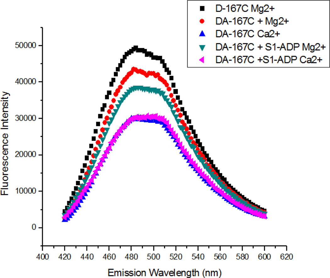
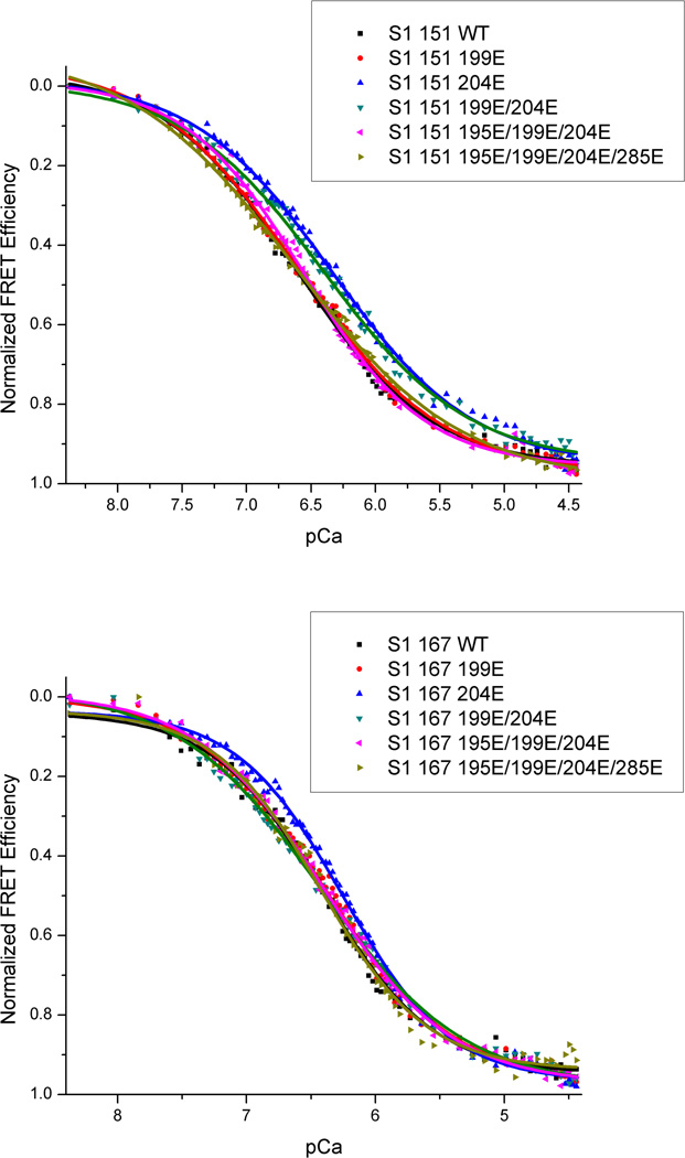
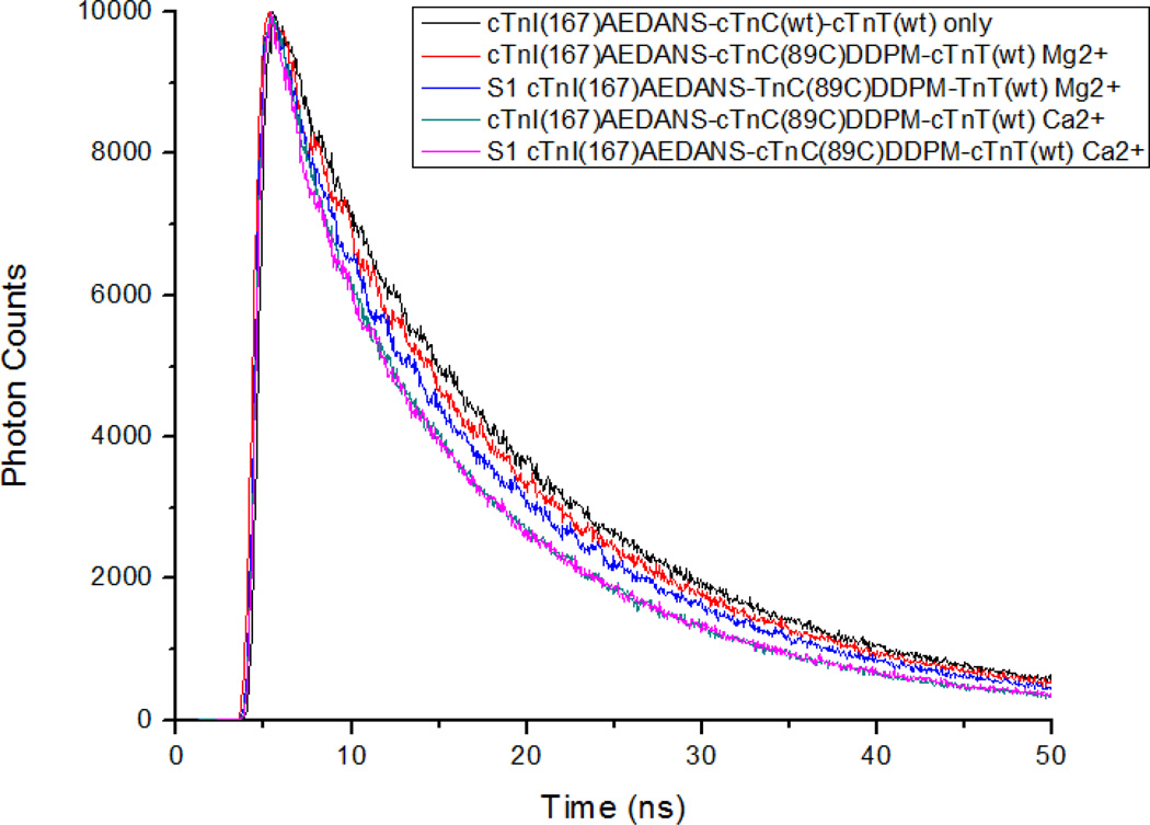
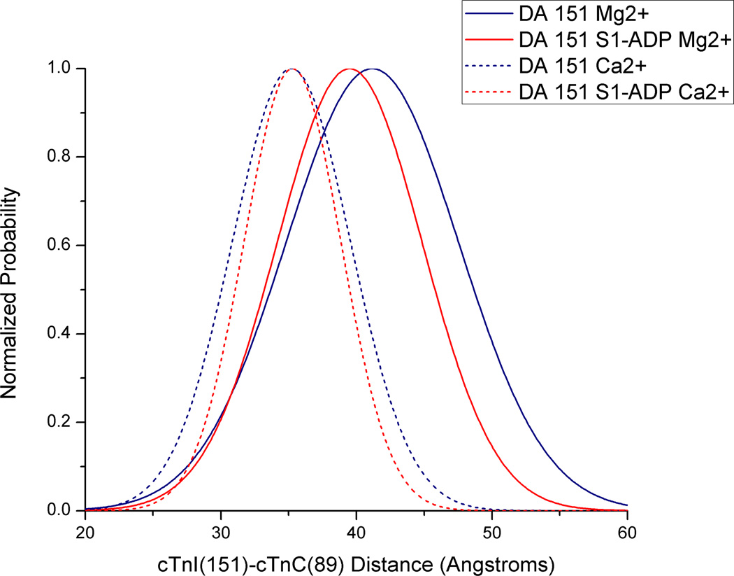
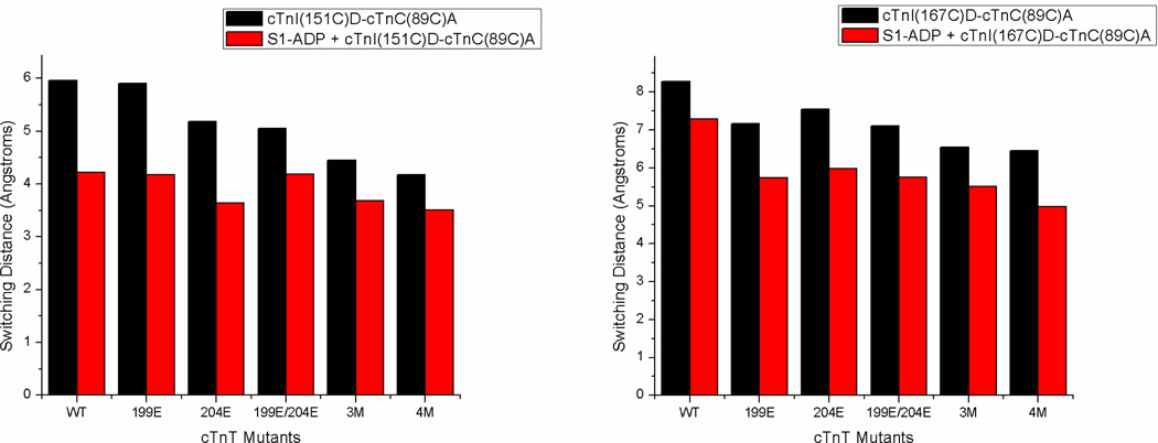
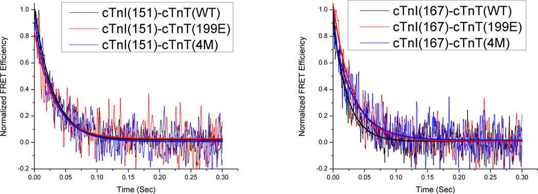
FRET was used to investigate the structural and kinetic effects that PKC phosphorylations exert on Ca(2+) and myosin subfragment-1 dependent conformational transitions of the cardiac thin filament. PKC phosphorylations of cTnT were mimicked by glutamate substitution. Ca(2+) and S1-induced distance changes between the central linker of cTnC and the switch region of cTnI (cTnI-Sr) were monitored in reconstituted thin filaments using steady state and time resolved FRET, while kinetics of structural transitions were determined using stopped flow. Thin filament Ca(2+) sensitivity was found to be significantly blunted by the presence of the cTnT(T204E) mutant, whereas pseudo-phosphorylation at additional sites increased the Ca(2+)-sensitivity. The rate of Ca(2+)-dissociation induced structural changes was decreased in the C-terminal end of cTnI-Sr in the presence of pseudo-phosphorylations while remaining unchanged at the N-terminal end of this region. Additionally, the distance between cTnI-Sr and cTnC was decreased significantly for the triple and quadruple phosphomimetic mutants cTnT(T195E/S199E/T204E) and cTnT(T195E/S199E/T204E/T285E), which correlated with the Ca(2+)-sensitivity increase seen in these same mutants. We conclude that significant changes in thin filament Ca(2+)-sensitivity, structure and kinetics are brought about through PKC phosphorylation of cTnT. These changes can either decrease or increase Ca(2+)-sensitivity and likely play an important role in cardiac regulation.
Keywords: Cardiac troponin T; FRET; PKC phosphorylation; Thin filament regulation.
Copyright © 2014 Elsevier Inc. All rights reserved.
Figures
Similar articles
-
Structural and kinetic effects of hypertrophic cardiomyopathy related mutations R146G/Q and R163W on the regulatory switching activity of rat cardiac troponin I.Arch Biochem Biophys. 2013 Jul 1;535(1):56-67. doi: 10.1016/j.abb.2012.12.007. Epub 2012 Dec 13. Arch Biochem Biophys. 2013. PMID: 23246786 Free PMC article.
-
Pathogenesis associated with a restrictive cardiomyopathy mutant in cardiac troponin T is due to reduced protein stability and greatly increased myofilament Ca2+ sensitivity.Biochim Biophys Acta. 2015 Feb;1850(2):365-72. doi: 10.1016/j.bbagen.2014.09.029. Epub 2014 Nov 1. Biochim Biophys Acta. 2015. PMID: 25450489 Free PMC article.
-
Effects of PKA phosphorylation of cardiac troponin I and strong crossbridge on conformational transitions of the N-domain of cardiac troponin C in regulated thin filaments.Biochemistry. 2007 Aug 28;46(34):9752-61. doi: 10.1021/bi700574n. Epub 2007 Aug 3. Biochemistry. 2007. PMID: 17676764 Free PMC article.
-
Molecular and integrated biology of thin filament protein phosphorylation in heart muscle.Ann N Y Acad Sci. 2004 May;1015:39-52. doi: 10.1196/annals.1302.004. Ann N Y Acad Sci. 2004. PMID: 15201148 Review.
-
Posttranslational modifications of cardiac troponin T: an overview.J Mol Cell Cardiol. 2013 Oct;63:47-56. doi: 10.1016/j.yjmcc.2013.07.004. Epub 2013 Jul 18. J Mol Cell Cardiol. 2013. PMID: 23871791 Review.
Cited by
-
Interplay Between the Effects of Dilated Cardiomyopathy Mutation (R206L) and the Protein Kinase C Phosphomimic (T204E) of Rat Cardiac Troponin T Are Differently Modulated by α- and β-Myosin Heavy Chain Isoforms.J Am Heart Assoc. 2016 Mar 21;5(3):e002777. doi: 10.1161/JAHA.115.002777. J Am Heart Assoc. 2016. PMID: 27001966 Free PMC article.
-
Stepwise C-Terminal Truncation of Cardiac Troponin T Alters Function at Low and Saturating Ca2.Biophys J. 2018 Aug 21;115(4):702-712. doi: 10.1016/j.bpj.2018.06.028. Epub 2018 Jul 12. Biophys J. 2018. PMID: 30057009 Free PMC article.
-
Fluorescence Based Characterization of Calcium Sensitizer Action on the Troponin Complex.Chem Biol Drug Des. 2016 Feb;87(2):171-81. doi: 10.1111/cbdd.12651. Epub 2015 Sep 16. Chem Biol Drug Des. 2016. PMID: 26375298 Free PMC article.
-
Pathomechanisms in heart failure: the contractile connection.J Muscle Res Cell Motil. 2015 Feb;36(1):47-60. doi: 10.1007/s10974-014-9395-8. Epub 2014 Nov 7. J Muscle Res Cell Motil. 2015. PMID: 25376563 Review.
-
Clinically Divergent Mutation Effects on the Structure and Function of the Human Cardiac Tropomyosin Overlap.Biochemistry. 2017 Jul 5;56(26):3403-3413. doi: 10.1021/acs.biochem.7b00266. Epub 2017 Jun 21. Biochemistry. 2017. PMID: 28603979 Free PMC article.
References
-
- Ebashi S, Endo M, Otsuki I. Q Rev Biophys. 1969;2:351–384. - PubMed
-
- Farah CS, Reinach FC. FASEB J. 1995;9:755–767. - PubMed
-
- Jin JP, editor. Nova Biomedical. Hauppauge, NY: 2013. Conformational states and behavior of the heterotrimeric troponin complex.
-
- Haselgrove JC. Cold Spring Harbor Symp. Qant. Biol. 1972;37:341–352.
-
- Huxley HE. Cold Spring Harbor Symp. Qant. Biol. 1972;37:361–376.
Publication types
MeSH terms
Substances
Grants and funding
LinkOut - more resources
Full Text Sources
Other Literature Sources
Molecular Biology Databases
Research Materials
Miscellaneous
