

A review on the wettability of dental implant surfaces II: Biological and clinical aspects
- PMID: 24709541
- PMCID: PMC4103435
- DOI: 10.1016/j.actbio.2014.03.032
A review on the wettability of dental implant surfaces II: Biological and clinical aspects
Abstract
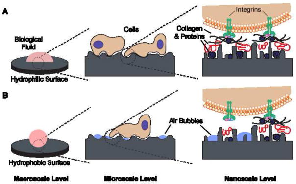
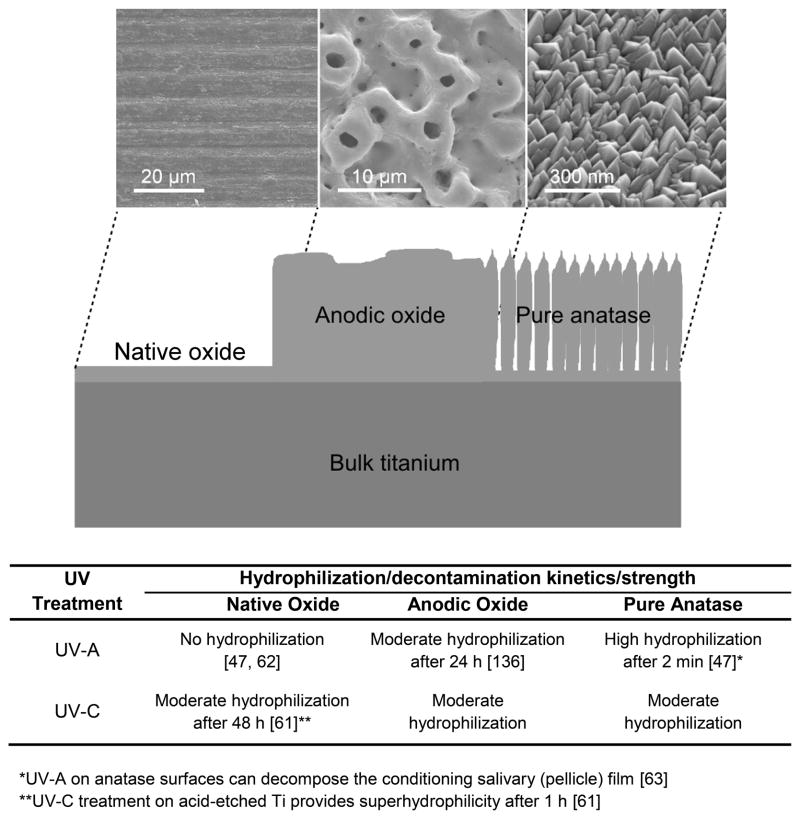
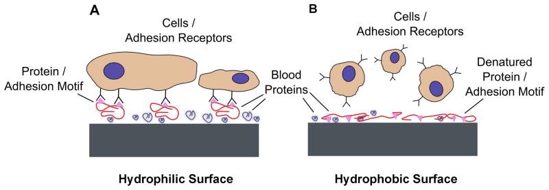
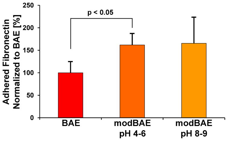
Dental and orthopedic implants have been under continuous advancement to improve their interactions with bone and ensure a successful outcome for patients. Surface characteristics such as surface topography and surface chemistry can serve as design tools to enhance the biological response around the implant, with in vitro, in vivo and clinical studies confirming their effects. However, the comprehensive design of implants to promote early and long-term osseointegration requires a better understanding of the role of surface wettability and the mechanisms by which it affects the surrounding biological environment. This review provides a general overview of the available information about the contact angle values of experimental and of marketed implant surfaces, some of the techniques used to modify surface wettability of implants, and results from in vitro and clinical studies. We aim to expand the current understanding on the role of wettability of metallic implants at their interface with blood and the biological milieu, as well as with bacteria, and hard and soft tissues.
Keywords: Hydrophilicity; Osseointegration; Surface conditioning; Surface energy; Titanium implant roughness.
Copyright © 2014 Acta Materialia Inc. Published by Elsevier Ltd. All rights reserved.
Figures
Similar articles
-
A review on the wettability of dental implant surfaces I: theoretical and experimental aspects.Acta Biomater. 2014 Jul;10(7):2894-906. doi: 10.1016/j.actbio.2014.02.040. Epub 2014 Feb 28. Acta Biomater. 2014. PMID: 24590162 Free PMC article. Review.
-
Surface characteristics of dental implants: A review.Dent Mater. 2018 Jan;34(1):40-57. doi: 10.1016/j.dental.2017.09.007. Epub 2017 Oct 10. Dent Mater. 2018. PMID: 29029850 Review.
-
Influence of Surface Contaminants and Hydrocarbon Pellicle on the Results of Wettability Measurements of Titanium.Int J Mol Sci. 2023 Sep 28;24(19):14688. doi: 10.3390/ijms241914688. Int J Mol Sci. 2023. PMID: 37834133 Free PMC article.
-
Hydrophilic implants generated using a low-cost dielectric barrier discharge plasma device at the time of placement exhibit increased osseointegration in an animal pre-clinical study: An effect that is sex-dependent.Dent Mater. 2022 Apr;38(4):632-645. doi: 10.1016/j.dental.2022.02.002. Epub 2022 Feb 18. Dent Mater. 2022. PMID: 35184898 Free PMC article.
-
Thermal-induced hydrophilicity enhancement of titanium dental implant surfaces.J Oral Sci. 2020 Mar 28;62(2):217-221. doi: 10.2334/josnusd.19-0235. Epub 2020 Mar 11. J Oral Sci. 2020. PMID: 32161230
Cited by
-
Polyether Ether Ketone Coated with Ultra-Thin Films of Titanium Oxide and Zirconium Oxide Fabricated by DC Magnetron Sputtering for Biomedical Application.Materials (Basel). 2022 Nov 14;15(22):8029. doi: 10.3390/ma15228029. Materials (Basel). 2022. PMID: 36431515 Free PMC article.
-
Gingival Response to Dental Implant: Comparison Study on the Effects of New Nanopored Laser-Treated vs. Traditional Healing Abutments.Int J Mol Sci. 2020 Aug 22;21(17):6056. doi: 10.3390/ijms21176056. Int J Mol Sci. 2020. PMID: 32842709 Free PMC article. Clinical Trial.
-
Osseointegration of a novel 3D porous Ti-6Al-4V implant material - Histomorphometric analysis in rabbits.Histol Histopathol. 2021 Aug;36(8):879-888. doi: 10.14670/HH-18-342. Epub 2021 May 11. Histol Histopathol. 2021. PMID: 33973645
-
Synergistic Enhancement of Protein Recruitment and Retention via Implant Surface Microtopography and Superhydrophilicity in a Computational Fluid Dynamics Model.Int J Mol Sci. 2023 Oct 26;24(21):15618. doi: 10.3390/ijms242115618. Int J Mol Sci. 2023. PMID: 37958605 Free PMC article.
-
Use of Piranha Solution as An Alternative Route to Promote Bioactivation of PEEK Surface with Low Functionalization Times.Molecules. 2022 Dec 22;28(1):74. doi: 10.3390/molecules28010074. Molecules. 2022. PMID: 36615270 Free PMC article.
References
-
- Karoussis IK, Bragger U, Salvi GE, Burgin W, Lang NP. Effect of implant design on survival and success rates of titanium oral implants: a 10-year prospective cohort study of the ITI Dental Implant System. Clin Oral Implants Res. 2004;15:8–17. - PubMed
-
- Albrektsson T, Zarb G, Worthington P, Eriksson AR. The long-term efficacy of currently used dental implants: a review and proposed criteria of success. Int J Oral Maxillofac Implants. 1986;1:11–25. - PubMed
-
- Fransson C, Wennstrom J, Berglundh T. Clinical characteristics at implants with a history of progressive bone loss. Clin Oral Implants Res. 2008;19:142–7. - PubMed
-
- Jacobs JJ, Andersson GBJ, Bell JE, Weinstein SL, Dormans JP, Gnatz SM, et al. United States Bone and Joint Decade: The burden of musculoskeletal diseases in the United States. 1. Rosemont: AAOS; 2008.
-
- Schwartz Z, Boyan BD. Underlying Mechanisms at the Bone-Biomaterial Interface. J Cell Biochem. 1994;56:340–7. - PubMed
Publication types
MeSH terms
Substances
Grants and funding
LinkOut - more resources
Full Text Sources
Other Literature Sources
Medical
