. 2013 Mar 14;2(1):163-87.
doi: 10.3390/cells2010163.

Regulation of Apoptosis by Inhibitors of Apoptosis (IAPs)

Affiliations

Regulation of Apoptosis by Inhibitors of Apoptosis (IAPs)

Jean Berthelet et al. Cells. .

Abstract

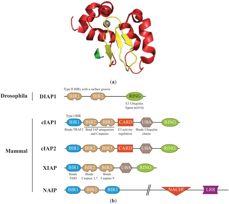
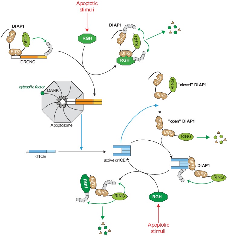
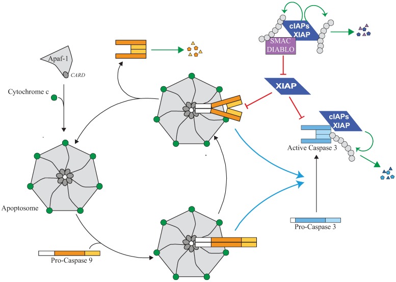
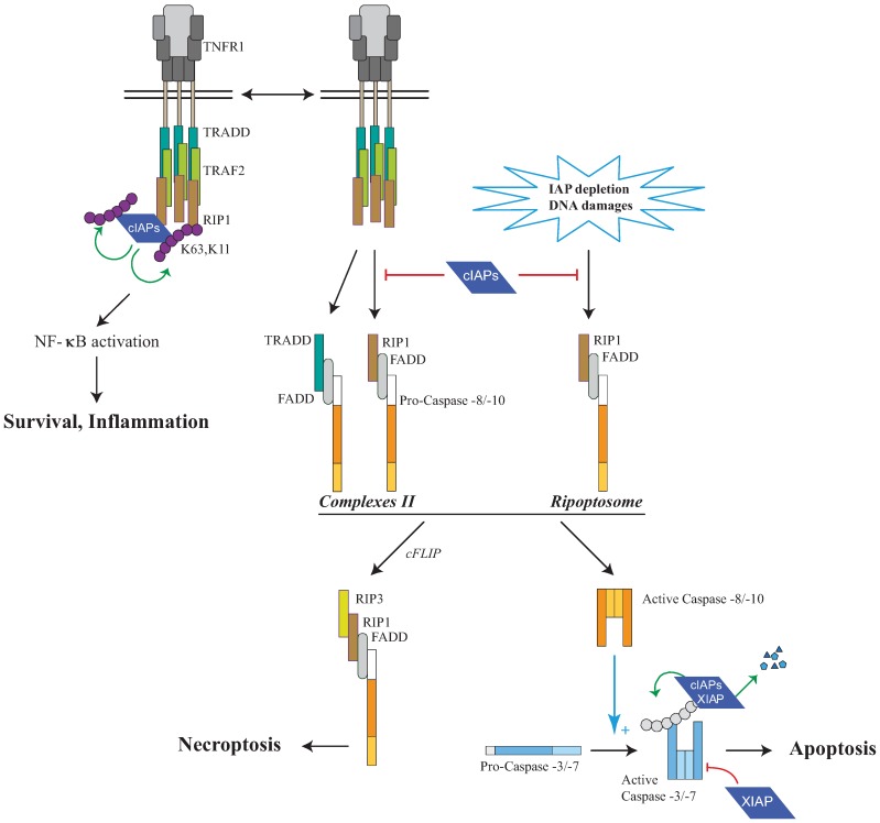
Inhibitors of Apoptosis (IAPs) are a family of proteins with various biological functions including regulation of innate immunity and inflammation, cell proliferation, cell migration and apoptosis. They are characterized by the presence of at least one N-terminal baculoviral IAP repeat (BIR) domain involved in protein-protein interaction. Most of them also contain a C-terminal RING domain conferring an E3-ubiquitin ligase activity. In drosophila, IAPs are essential to ensure cell survival, preventing the uncontrolled activation of the apoptotic protease caspases. In mammals, IAPs can also regulate apoptosis through controlling caspase activity and caspase-activating platform formation. Mammalian IAPs, mainly X-linked IAP (XIAP) and cellular IAPs (cIAPs) appeared to be important determinants of the response of cells to endogenous or exogenous cellular injuries, able to convert the survival signal into a cell death-inducing signal. This review highlights the role of IAP in regulating apoptosis in Drosophila and Mammals.


Figures

Figure 1
Figure 1
Structure of the Inhibitors of apoptosis. (a) Structure of the cellular IAP1 (cIAP1)-baculoviral IAP repeat (BIR)3 bound to the caspase-9 N-terminal peptide [30]. BIR3 is organized in four α-helices (red) and 3 β-strand sheets (yellow) maintained by zinc ion (grey). The interaction involved the surface hydrophobic groove of cIAP1 and the N-terminal peptide (ATPFQ) of the caspase 9 sub-unit (Constructed using the PyMOL Molecular Graphics System). (b)Representation of IAPs involved in the regulation of apoptosis. The type I baculoviral IAP repeats (BIR, blue) of cIAPs and X-linked IAP (XIAP) can bind to cell signalling intermediates TNFR associated factor 2 (TRAF2) and Transforming Growth Factor beta-activated kinase 1-binding protein 1 (TAB1), respectively. The type II BIRs (brown) contain a surface hydrophobic groove allowing the interaction with IBM found in caspase sub-units and IAP antagonists. The ubiquitin Associated (UBA) domain binds ubiquitin chains. The caspase recruitment domain (CARD) is a module of regulation of the RING E3-ubiquitin ligase activity. The RING domain confers to IAPs an E3-ubiquitin ligase activity. NACHT (domain present in NAIP, CIITA, HETE and TP1). LRR: Leucine Rich Repeat.
Figure 2
Figure 2
Regulation of the caspase cascade by IAPs in drosophila. In living cells, the caspase activating cascade is maintained in check by a direct interaction of caspases with the Drosophila IAP1 (DIAP1). The DIAP1 BIR2 binds to the prodomain of the apoptotic initiator DROsophila Nedd-2-like Caspase (DRONC) and the RING induces DRONC ubiquitination preventing apoptosome assembly. DIAP1 is expressed in “closed conformation” in which the N-terminal sequence hides BIR1 surface groove. Effector caspase mediates the cleavage of the N-extremity of DIAP1 that releases the BIR1 domain which in turn interacts with the IBM exposed on the active form of effector drosophila melanogaster Interleukin-1-converting enzyme/Ced-3 related protease (drICE). DIAP1 inhibits drICE activity through a degradative or non-degradative ubiquitination or neddylation. The “open” form of DIAP1 is highly unstable and rapidly degraded by the N-end-rule-associated degradation machinery. Apoptotic stimuli induce the expression of the IBM-carrying IAP antagonists Reaper, Grim or Hid (RGH) wich strongly bind and neutralize DIAP1 through IBM-BIRs interaction. The IAP antagonist-DIAP1 interaction promotes DIAP1-autoubiquitination and degradation.
Figure 3
Figure 3
Regulation of the apoptosome and caspase activity by IAPs. The release of cytochrome-c from mitochondria which occurs during intrinsic pathway of apoptosis triggers an ATP-dependent conformational change and oligomerisation of the adaptor apoptotic protease activating factor 1 (Apaf-1) in a heptameric complex apoptosome. Apaf-1 then recruits Caspase-9 via its pro-domain through a homotypic CARD-CARD interaction. Caspase-9 is activated by homodimerisation and promotes the activating cleavage of effector caspase-3 leading to apoptosis. Caspase-9 undergoes autocatalytic processing and is then quickly disconnected from the apoptosome which is free to recruit a new pro-caspase-9. XIAP can control caspase activating pathway at several steps. First, XIAP is present in the apoptosome where it directly binds processed caspase-9 and inhibits its activity. The inhibition of caspase-9 by XIAP could stabilize the caspase-9 apoptosome complex and block the cycle of caspase-9 activation. Second, XIAP can directly bind and inhibit active effector caspase-3. XIAP can inhibit caspase activity by hindering substrate accessibility or hiding the protease catalytic residue, and/or by promoting ubiquitination or neddylation. Although unable to inhibit their activity, cIAP1 can bind to processed caspases and promote their ubiquitination. An IBM-dependent binding of IAP antagonists such as Smac/Diablo prevents XIAP-mediated caspase inhibition while cIAPs could interfere with neutralizing binding XIAP-Smac/Diablo.
Figure 4
Figure 4
Regulation of RIP1-containing platforms by IAPs. Tumor Necrosis Factor Receptor 1 (TNFR1) stimulation induces the recruitment, to the receptor, of cIAPs and Receptor interacting protein 1 (RIP1) via the adaptors TNFR1-associated Death domain (TRADD) and TNFR associated factor (TRAF2). cIAPs trigger K63 self-ubiquitination and K11 and K63-ubiquitination of RIP1 leading to NF-κB activation and survival. In the absence of cIAPs, a secondary RIP1-containing cytoplasmic complex is formed (complex II) leading to cell death. A cytoplasmic RIP-1-containing complex named RIPoptosome can also be assembled, in the absence of Death Receptor stimulation, after DNA-damage-mediated IAP depletion or the use of synthetic IAP antagonists known to induce IAP degradation. RIP1-containing platform can lead to either caspase-8 or -10 activation and apoptosis, or caspase-independent cell death referred to as Necroptosis. Initiator caspases-8 or -10 induce the activating proteolytic processing of effector caspase-3 or -7 responsible for apoptosis. IAPs can control cell death at different levels: (1) cIAPs can induce the K63 and K11 ubiquitination of RIP1 allowing NF-κB activation and preventing the formation of complex II or RIPoptosome; (2) cIAPs and XIAP can induce K48-ubiquitination of RIP1 leading to its proteasomal degradation. (3) XIAP can directly inhibit the activity of processed effector caspase-3 or -7; (4) cIAPs and XIAP can induce ubiquitination and proteasome-mediated degradation of processed forms of caspase-3 or -7.

References

    1. Clarke T.E., Clem R.J. Insect defenses against virus infection: The role of apoptosis. Int. Rev. Immunol. 2003;22:401–424. doi: 10.1080/08830180305215. - DOI - PubMed
    1. Crook N.E., Clem R.J., Miller L.K. An apoptosis-inhibiting baculovirus gene with a zinc finger-like motif. J. Virol. 1993;67:2168–2174. - PMC - PubMed
    1. Orme M., Meier P. Inhibitor of apoptosis proteins in Drosophila: Gatekeepers of death. Apoptosis. 2009;14:950–960. doi: 10.1007/s10495-009-0358-2. - DOI - PubMed
    1. Marivin A., Berthelet J., Plenchette S., Dubrez L. The inhibitor of apoptosis (IAPs) in adaptive response to cellular stress. Cells. 2012;1:711–737. doi: 10.3390/cells1040711. - DOI - PMC - PubMed
    1. Beug S.T., Cheung H.H., Lacasse E.C., Korneluk R.G. Modulation of immune signalling by inhibitors of apoptosis. Trends Immunol. 2012;33:535–545. doi: 10.1016/j.it.2012.06.004. - DOI - PubMed

LinkOut - more resources