. 2014 Jun 1;277(2):118-23.
doi: 10.1016/j.taap.2014.03.017. Epub 2014 Apr 4.

Aspirin-triggered resolvin D1 down-regulates inflammatory responses and protects against endotoxin-induced acute kidney injury

Affiliations

Aspirin-triggered resolvin D1 down-regulates inflammatory responses and protects against endotoxin-induced acute kidney injury

Jiao Chen et al. Toxicol Appl Pharmacol. .

Abstract

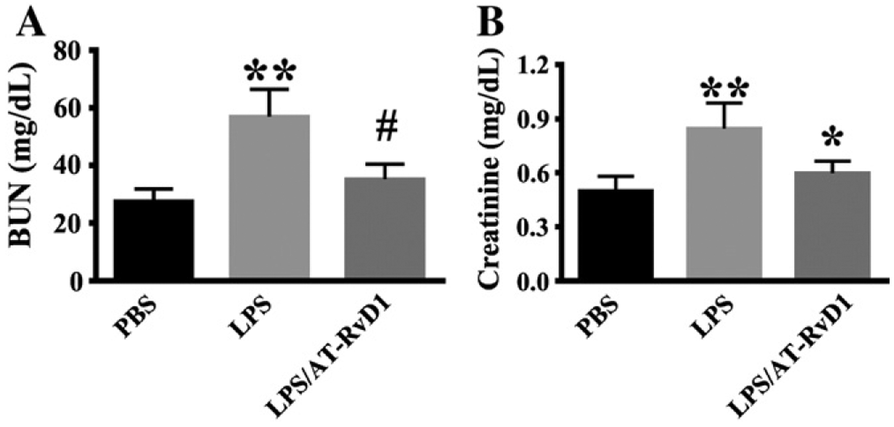
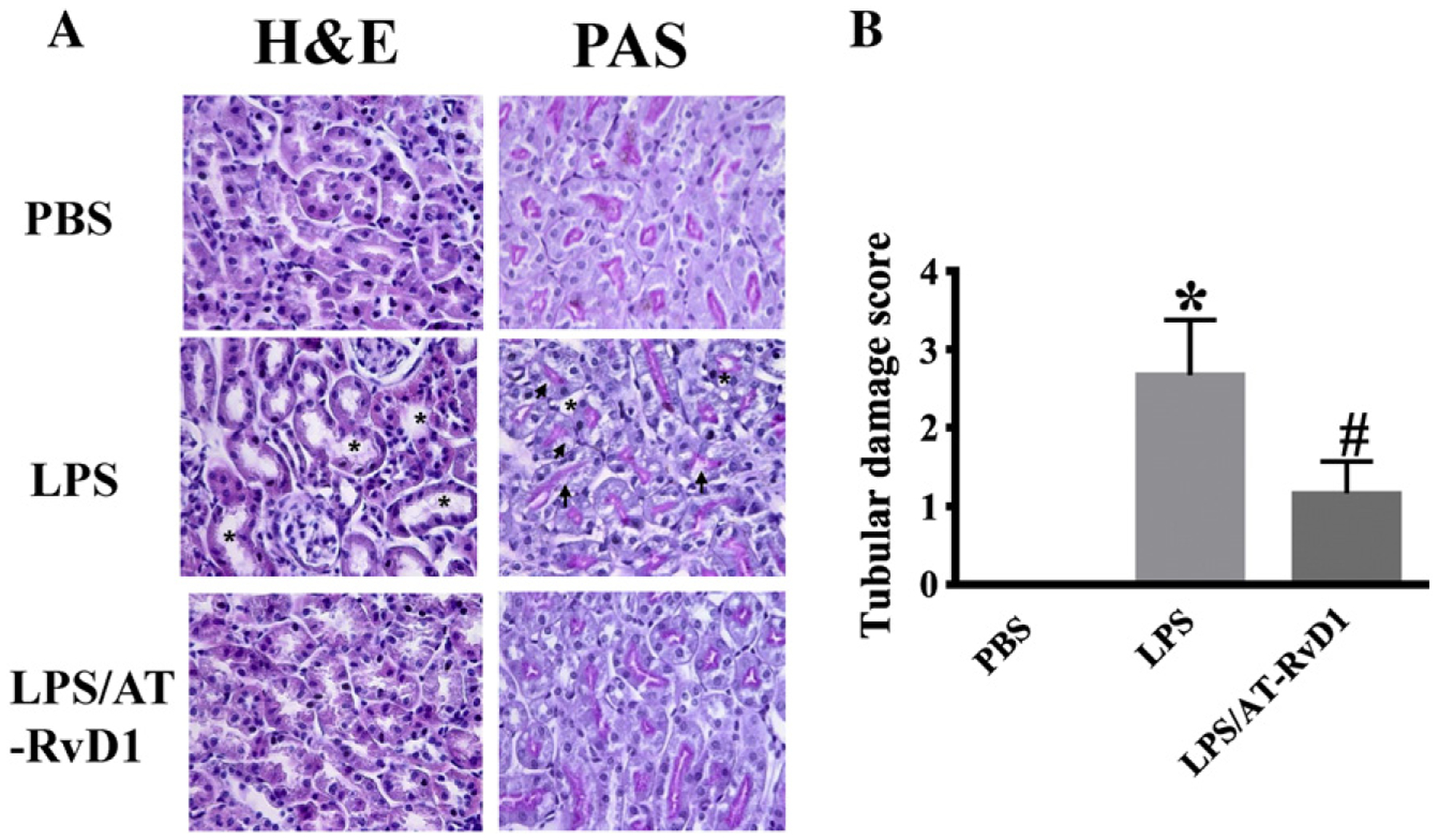
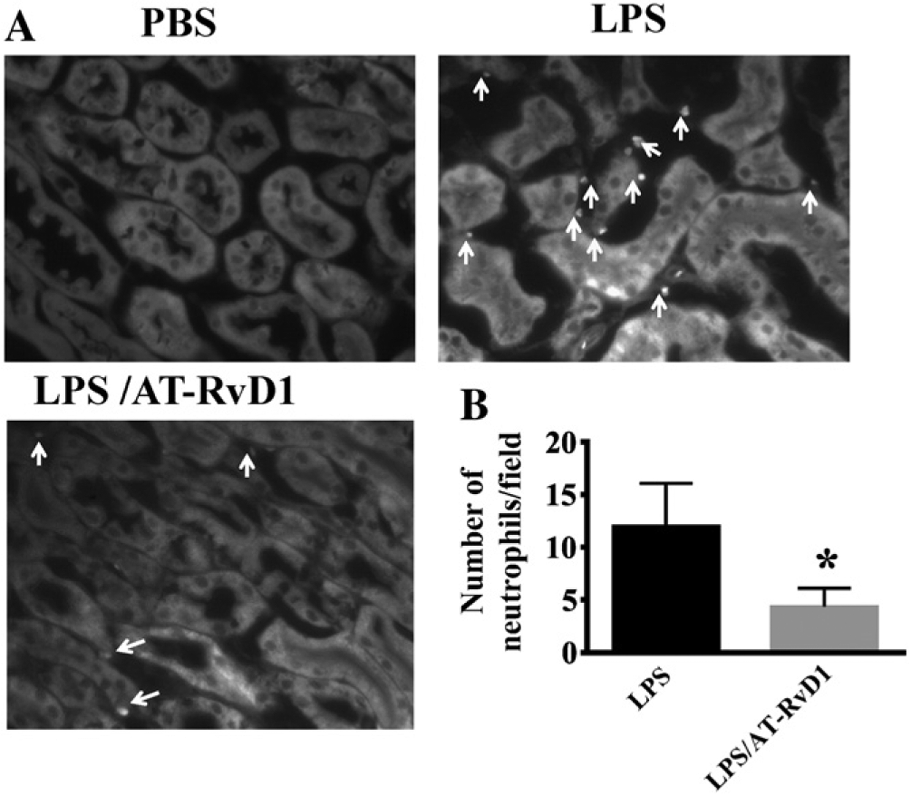
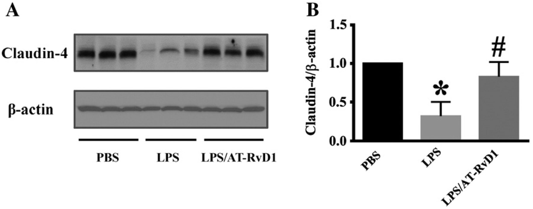
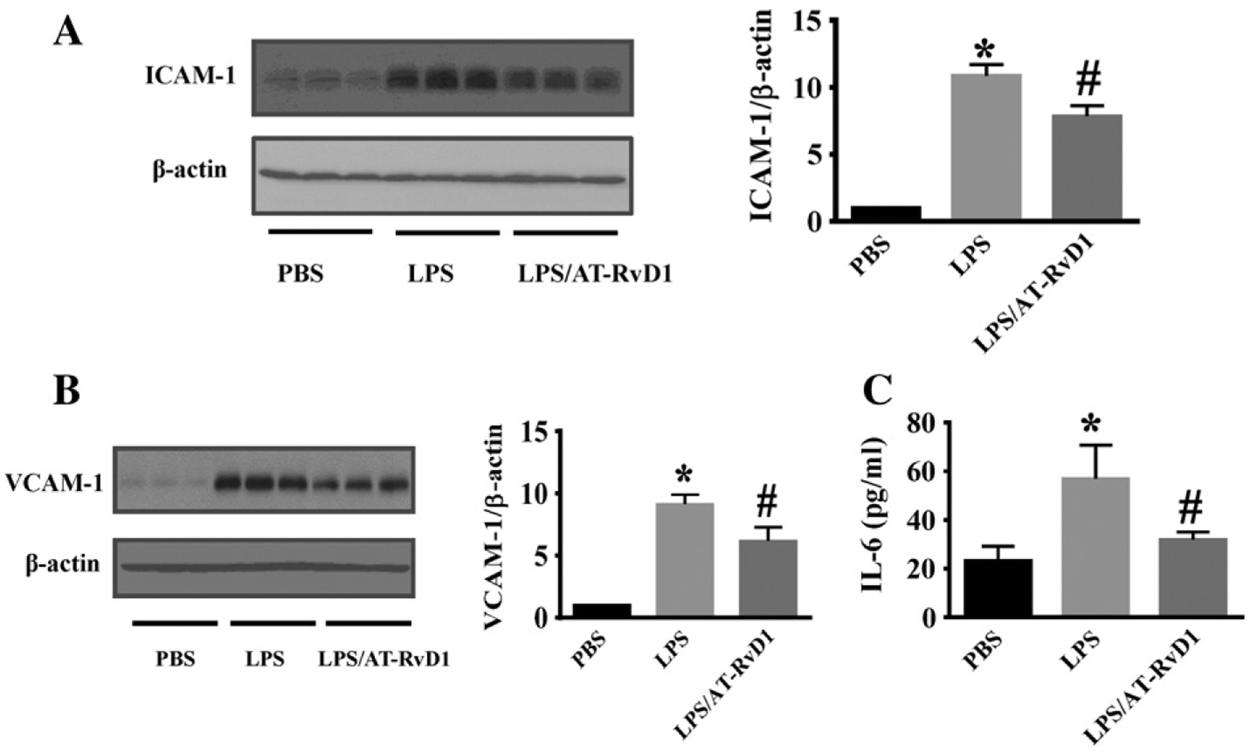
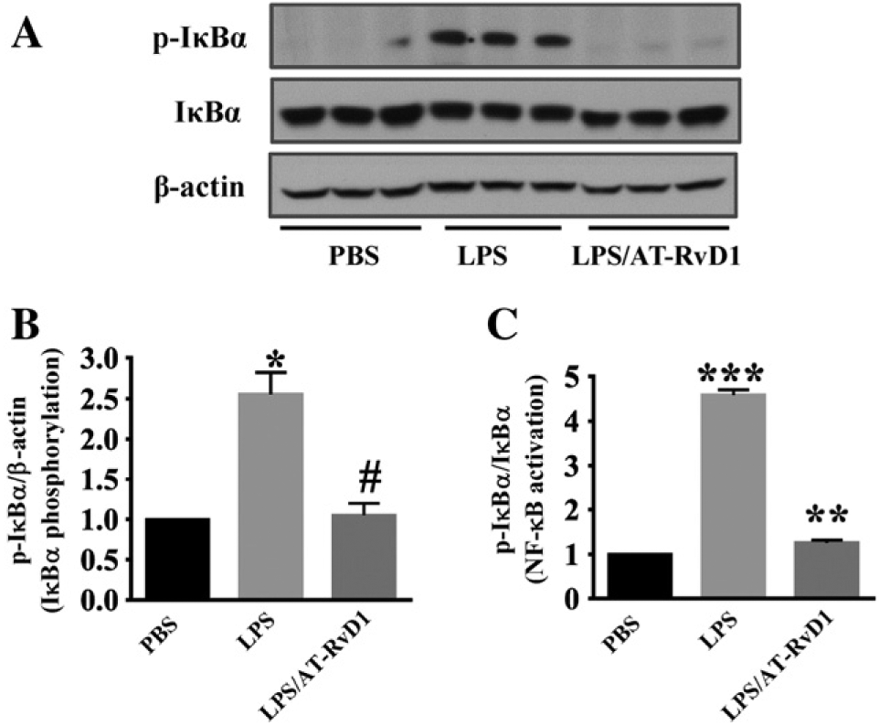
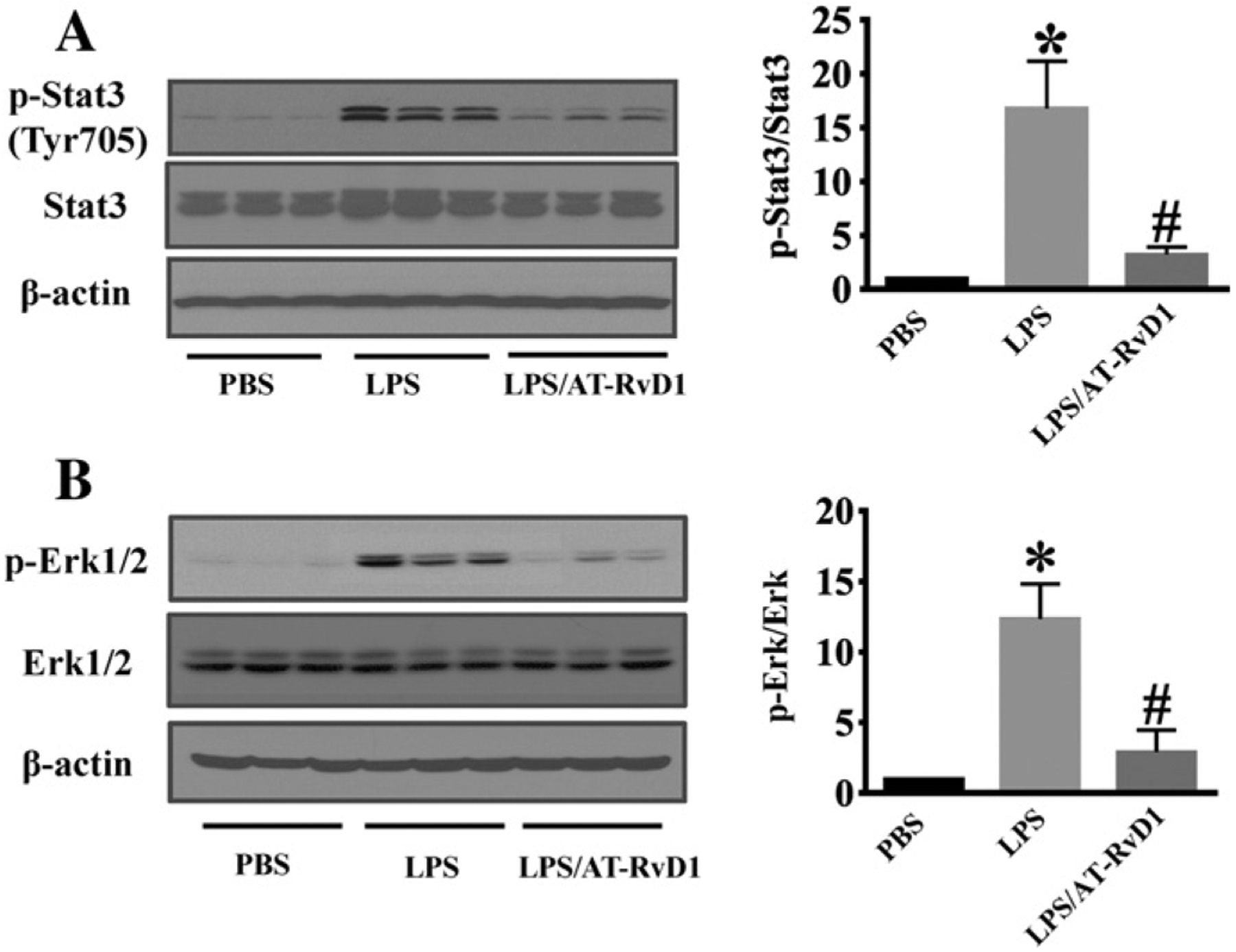
The presence of endotoxin in blood can lead to acute kidney injury (AKI) and septic shock. Resolvins, the endogenous lipid mediators derived from docosahexaenoic acid, have been reported to exhibit potent anti-inflammatory action. Using a mouse model of lipopolysaccharide (LPS)-induced AKI, we investigated the effects of aspirin-triggered resolvin D1 (AT-RvD1) on inflammatory kidney injury. Administration of AT-RvD1 1h after LPS challenge protected the mice from kidney injury as indicated by the measurements of blood urea nitrogen, serum creatinine, and morphological alterations associated with tubular damage. The protective effects were evidenced by decreased neutrophil infiltration in the kidney indicating reduction in inflammation. AT-RvD1 treatment restored kidney cell junction protein claudin-4 expression, which was otherwise reduced after LPS challenge. AT-RvD1 treatment inhibited endotoxin-induced NF-κB activation and suppressed LPS-induced ICAM-1 and VCAM-1 expression in the kidney. Moreover, AT-RvD1 treatment markedly decreased LPS-induced IL-6 level in the kidney and blocked IL-6-mediated signaling including STAT3 and ERK phosphorylation. Our findings demonstrate that AT-RvD1 is a potent anti-inflammatory mediator in LPS-induced kidney injury, and AT-RvD1 has therapeutic potential against AKI during endotoxemia.

Keywords: Acute kidney injury; Aspirin-triggered resolvin D1; Endotoxemia; Inflammation; Sepsis.


Conflict of interest statement

Disclosure

All the authors declared no competing interests.

Figures

Fig. 1.
Fig. 1.
AT-RvD1 protects against renal dysfunction during LPS-induced kidney injury. Mice were divided into three groups: PBS control (PBS); 5 mg/Kg LPS (LPS); 5 mg/kg LPS/1 μg AT-RvD1 (LPS/AT-RvD1). Mice were treated with AT-RvD1 (1 μg) or PBS at 1 h after LPS challenge. (A) Blood urea nitrogen levels in each group at 24 h after LPS (PBS: n = 9 mice; LPS: n = 10 mice; LPS/AT-RvD1: n = 7 mice). (B) Serum creatinine levels in each group at 24 h after LPS challenge (PBS: n = 5 mice; LPS: n = 10 mice; LPS/AT-RvD1: n = 8 mice). Data are presented as mean ± SD. **P < 0.0001 vs PBS group; #P < 0.0001 vs LPS group; *P < 0.001 vs LPS group.
Fig. 2.
Fig. 2.
AT-RvD1 protects against LPS-induced renal tubular damage. Kidney tissues were collected at 24 h after LPS challenge for H&E and PAS staining (PBS: n = 6 mice; LPS: n = 9 mice; LPS/AT-RvD1: n = 6 mice). (A) Representative H&E staining (A) and PAS staining (B) of kidney cortex and outer medulla tissues. Compared with WT mice, LPS-induced renal tubular injury with tubular dilation or cast formation (*) and vacuolization of renal tubular cells (arrows). (C) Pathological scores of tubular damage in LPS and LPS/AT-RvD1 groups. Data are presented as mean ± SD. *P < 0.0001 vs PBS group, #P < 0.001 vs LPS group.
Fig. 3.
Fig. 3.
AT-RvD1 inhibits neutrophil infiltration in LPS-induced acute kidney injury. Kidney sections were stained with FITC-anti-GR1 antibody specific for neutrophils. (A) The outer medullary region of sections was photographed at ×200. White arrows indicate neutrophils. (B) Neutrophil count/field (LPS: n = 8 mice; LPS/AT-RvD1: n = 6 mice). *P < 0.001 vs LPS group.
Fig. 4.
Fig. 4.
AT-RvD1 regulates the cell junction protein claudin-4 expression in acute kidney injury. Kidney tissues were subjected to immunoblotting assay (PBS: n = 8 mice; LPS: n = 10 mice; LPS/AT-RvD1: n = 9 mice). (A) Claudin-4 expression in kidney tissues. Blots were re-probed for actin to normalize total protein content. (B) Data are expressed as the relative ratio to actin (right panel), and presented as mean ± SD. *P < 0.05 vs PBS group; #P < 0.05 vs LPS group.
Fig. 5.
Fig. 5.
AT-RvD1 inhibits LPS-induced kidney inflammation. Kidney tissues were subjected to immunoblotting assay to assess ICAM-1 (A) and VCAM-1 (B) expression (PBS: n = 8 mice; LPS: n = 10 mice; LPS/AT-RvD1: n = 9 mice). Immunoblots were re-probed for actin to normalize total protein content. Data are expressed as the relative ratio to actin and presented as mean ± SD. *P < 0.0001, vs PBS group; #P < 0.05, vs LPS group. (C) Kidney IL-6 levels were determined by ELISA (n = 5 mice/group). Data are presented as mean ± SD. *P < 0.001 vs PBS group; #P < 0.05 vs LPS group.
Fig. 6.
Fig. 6.
AT-RvD1 inhibits NF-κB activation in LPS-induced acute kidney injury. Immunoblotting assays will conducted to examine phosphorylated IκBα, total IκBα and actin levels in kidney tissues (n = 6 mice/group). (A) Representative blots of phosphorylated IκBα, IκBα and actin. (B, C) Densitometry analysis is presented as relative ratios of phosphorylated IκBα to actin and total IκBα. Data are presented as mean ± SD. *P < 0.05 vs PBS group; #P < 0.05 vs LPS group. ***P < 0.0001 vs PBS group; **P < 0.001 vs LPS group.
Fig. 7.
Fig. 7.
AT-RvD1 inhibits inflammatory signaling during LPS-acute kidney injury. Kidney tissues was collected for immunoblotting assays (n = 6 mice/group). (A) Representative blots of LPS-induced STAT3 phosphorylation. (B) Representative blots of LPS-induced ERK phosphorylation. Densitometry analysis is presented as the relative ratio of each protein to non-phosphorylated proteins. Data are presented as mean ± SD. *P < 0.05 vs PBS group; #P < 0.05 vs LPS group.

References

    1. Amasheh S, Fromm M, Gunzel D, 2011. Claudins of intestine and nephron—a correlation of molecular tight junction structure and barrier function. Acta Physiol 201, 133–140. - PubMed
    1. Angus DC, Linde-Zwirble WT, Lidicker J, Clermont G, Carcillo J, Pinsky MR, 2001. Epidemiology of severe sepsis in the United States: analysis of incidence, outcome, and associated costs of care. Crit. Care Med 29, 1303–1310. - PubMed
    1. Bagshaw SM, Laupland KB, Doig CJ, Mortis G, Fick GH, Mucenski M, Godinez-Luna T, Svenson LW, Rosenal T, 2005. Prognosis for long-term survival and renal recovery in critically ill patients with severe acute renal failure: a population-based study. Crit. Care 9, R700–R709. - PMC - PubMed
    1. Balkovetz DF, 2006. Claudins at the gate: determinants of renal epithelial tight junction paracellular permeability. Am. J. Physiol. Ren. Physiol 290, F572–F579. - PubMed
    1. Bento AF, Claudino RF, Dutra RC, Marcon R, Calixto JB, 2011. Omega-3 fatty acidderived mediators 17(R)-hydroxy docosahexaenoic acid, aspirin-triggered resolvin D1 and resolvin D2 prevent experimental colitis in mice. J. Immunol 187, 1957–1969. - PubMed

MeSH terms