. 2014 Apr 7;9(4):e93991.
doi: 10.1371/journal.pone.0093991. eCollection 2014.

WRN loss induces switching of telomerase-independent mechanisms of telomere elongation

Affiliations

WRN loss induces switching of telomerase-independent mechanisms of telomere elongation

April Renee Sandy Gocha et al. PLoS One. .

Abstract

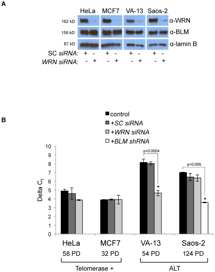
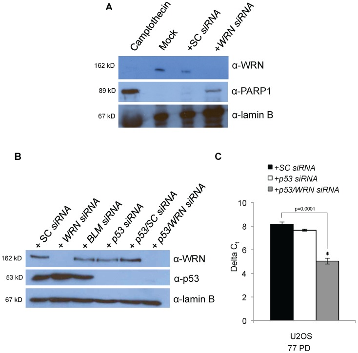
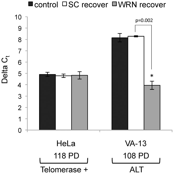
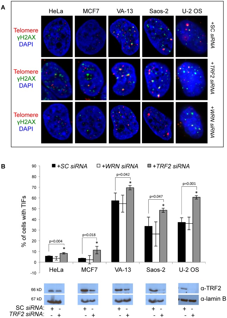
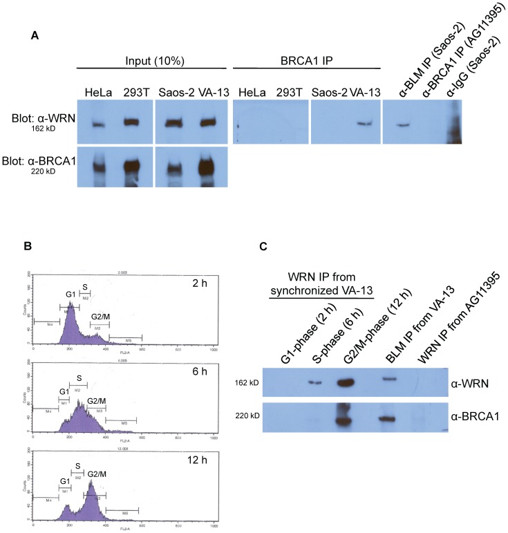
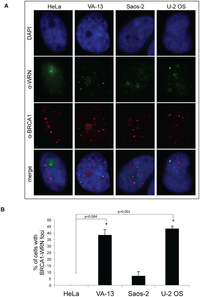
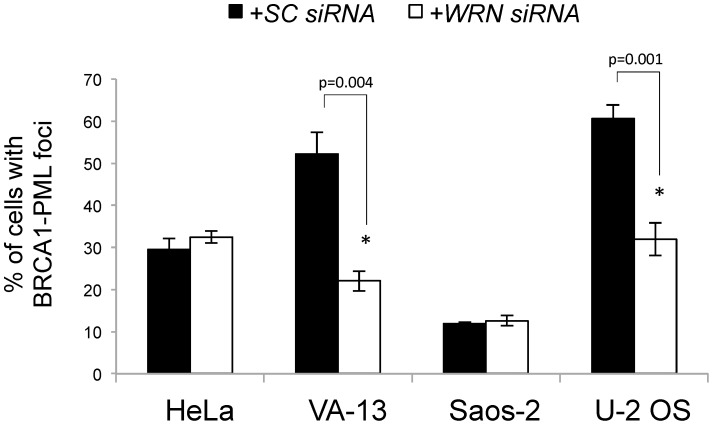
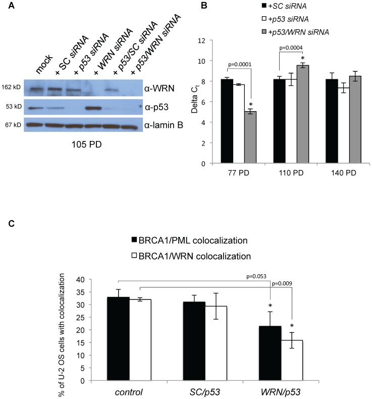
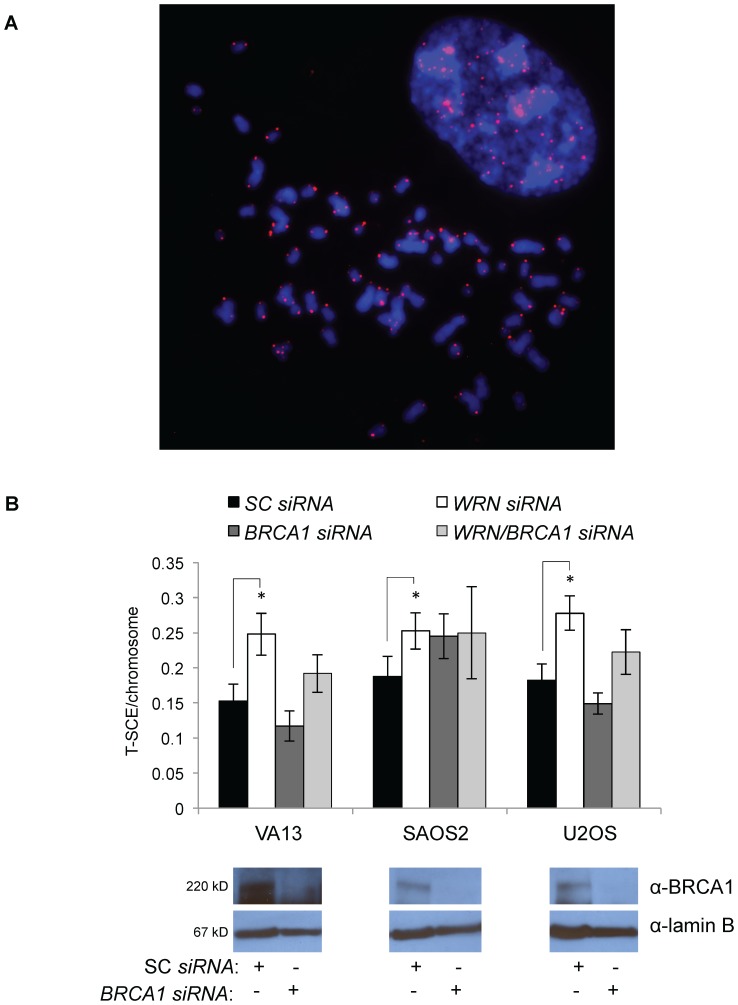
Telomere maintenance can occur in the presence of telomerase or in its absence, termed alternative lengthening of telomeres (ALT). ALT adds telomere repeats using recombination-based processes and DNA repair proteins that function in homologous recombination. Our previous work reported that the RecQ-like BLM helicase is required for ALT and that it unwinds telomeric substrates in vitro. WRN is also a RecQ-like helicase that shares many biochemical functions with BLM. WRN interacts with BLM, unwinds telomeric substrates, and co-localizes to ALT-associated PML bodies (APBs), suggesting that it may also be required for ALT processes. Using long-term siRNA knockdown of WRN in three ALT cell lines, we show that some, but not all, cell lines require WRN for telomere maintenance. VA-13 cells require WRN to prevent telomere loss and for the formation of APBs; Saos-2 cells do not. A third ALT cell line, U-2 OS, requires WRN for APB formation, however WRN loss results in p53-mediated apoptosis. In the absence of WRN and p53, U-2 OS cells undergo telomere loss for an intermediate number of population doublings (50-70), at which point they maintain telomere length even with the continued loss of WRN. WRN and the tumor suppressor BRCA1 co-localize to APBs in VA-13 and U-2 OS, but not in Saos-2 cells. WRN loss in U-2 OS is associated with a loss of BRCA1 from APBs. While the loss of WRN significantly increases telomere sister chromatid exchanges (T-SCE) in these three ALT cell lines, loss of both BRCA1 and WRN does not significantly alter T-SCE. This work demonstrates that ALT cell lines use different telomerase-independent maintenance mechanisms that variably require the WRN helicase and that some cells can switch from one mechanism to another that permits telomere elongation in the absence of WRN. Our data suggest that BRCA1 localization may define these mechanisms.


Conflict of interest statement

Competing Interests: The authors have declared that no competing interests exist.

Figures

Figure 1
Figure 1. WRN siRNA knockdown and telomere length of HeLa, MCF7, VA-13, and Saos-2 cells.
A. Pooled WRN siRNAs or scrambled control (SC) siRNAs were transfected into immortalized human cell lines and whole cell extracts were collected 48 hours after transfection. Each lysate was separated by SDS-PAGE and western blotted with antibodies to WRN (top), BLM (to ensure specificity of the siRNAs, middle), and lamin B (as a loading control, bottom). B. Relative telomere length measurement by qRT-PCR. The difference in the cycle threshold (Ct) between a telomere-specific PCR reaction and a single copy gene (ALB) PCR reaction is calculated for each sample as the ΔCt, which represents the average relative telomere repeat length. Telomere length in Saos-2 ALT cells decreases after stable BLM knockdown as measured by TRF Southern blot . Here, we confirm detection of these length changes by qRT-PCR (far right). Telomere lengths of telomerase-positive cells, HeLa and MCF7, and ALT cells, VA-13 and Saos2, were measured following transfection of either SC or WRN siRNAs. Relative lengths depicted represent the final measurement for each cell type, which was taken at the population doubling number (PD) indicated. (ALT = alternative lengthening of telomeres; TRF =  telomere restriction fragment; PD = population doubling).
Figure 2
Figure 2. WRN siRNA knockdown and telomere length of U-2 OS cells.
A. U-2 OS cells were mock transfected or transfected with SC or WRN siRNAs. Whole cell extracts were collected, separated by SDS-PAGE and western blotted with antibodies to WRN to confirm the knockdown (top panel), cleaved PARP1 to examine apoptosis induction (middle panel) and lamin B as a loading control (bottom panel). Treatment with camptothecin is a positive control for the induction of apoptosis. B. To inhibit apoptosis, U-2 OS cells were transfected with WRN and p53 siRNAs. Whole cell extracts were separated by SDS-PAGE and western blotted with antibodies specific to WRN (top panel), p53 (middle panel) and lamin B (loading control; bottom panel). C. DNA from transfected U-2 OS cells was collected at 77 PD and telomere length was measured by qRT-PCR. Average relative telomere repeat lengths are graphically represented as the difference between a telomere-specific PCR reaction in comparison to the single copy gene ALB PCR reaction, or delta Ct. (ALT = alternative lengthening of telomeres; PD = population doubling)
Figure 3
Figure 3. ALT-associated PML body (APB) formation following WRN knockdown.
A. Representative confocal micrograph images of cells from two telomerase-positive cell lines, HeLa and MCF7, and three ALT cell lines, VA-13, Saos-2 and U-2 OS. Cells were fixed 48 hours after transfection with scrambled control (SC) siRNAs, BLM siRNAs or WRN siRNAs and immunofluorescently labeled with antibodies to PML and TRF2. We have previously shown a reduction in APBs following BLM knockdown , so cells transfected with BLM siRNAs served as a positive control. PML is labeled in red, TRF2 in green, and nuclei stained with DAPI (blue). B. A graphical representation of at least 3 independent experiments depicts the percentage of cells displaying co-localization of TRF2 and PML, indicative of APBs. Co-localization of PML/TRF2 foci was scored as a positive indication of APBs; at least three independent experiments were averaged to yield the percentage of each cell type. Western blots confirm the ability of BLM siRNAs to reduce BLM expression.
Figure 4
Figure 4. Telomere length after recovery from WRN siRNA transfection.
After 54 PD of continuous WRN knockdown (after telomeres had shortened by almost 50% of the original length), WRN siRNA transfections of a subset of HeLa and VA-13 cells were ceased and cells allowed to recover in culture for an additional 54 PD. Average relative telomere repeat lengths were then measured by qRT-PCR. Results are graphically represented as the difference between a telomere-specific PCR reaction in comparison to the single copy gene albumin PCR reaction as delta Ct.
Figure 5
Figure 5. Telomere dysfunction-induced foci (TIF) formation after WRN knockdown.
A. Representative confocal micrograph images of cells from two telomerase positive cell lines, HeLa and MCF7, and three ALT cell lines, VA-13, Saos-2 and U-2 OS. Cells were fixed 48 hours after transfection with scrambled control (SC) siRNAs, WRN siRNAs or TRF2 siRNAs and subjected to fluorescent in situ hybridization (FISH) with a Cy3-labeled telomeric PNA probe and immunofluorescently labeled with an antibody to phosphorylated histone 2A (γH2AX). As TRF2 is a member of the shelterin complex, TRF2 knockdown was used as a positive control. The telomere is labeled with a Cy3-PNA probe in red, γH2AX in green, and the nucleus is stained with DAPI (blue). B. A graphical representation of at least three independent experiments were averaged to yield the percentage of each cell type with γH2AX co-localization with the telomere, indicative of telomere dysfunction induced foci (TIF). Western blots confirm the ability of TRF2 siRNAs to reduce TRF2 expression.
Figure 6
Figure 6. WRN immunoprecipitation from telomerase-positive and ALT cells.
A. Nuclear extracts were immunoprecipitated with antibodies to BRCA1 and the immunoprecipitated proteins subjected to western blotting with an antibody to WRN (right panels). Immunoprecipitation with anti-BLM serves as a positive control for the interaction with WRN in Saos-2 cell extracts, while an anti-BRCA1 immunoprecipitation from AG11395 WS cell extracts and an anti-IgG immunoprecipitation from Saos-2 cell extracts serve as negative controls. Immunoprecipitation input lanes (10% of the total extract) of WRN and BRCA1 are shown in the left panels. B. VA-13 cells were synchronized with aphidicolin and collected at various time points post-release from aphidicolin arrest. Cell cycle distribution was analyzed by flow cytometry at 2, 6 or 12 hours post-release as depicted in the histograms. C. Synchronized nuclear extracts were immunoprecipitated with antibodies to WRN and the immunoprecipitated proteins subjected to western blotting with antibodies to WRN (top) or BRCA1 (bottom). Immunoprecipitation with anti-BLM serves as a positive control for the interaction with WRN and BRCA1 in VA-13 cell extracts, while an anti-WRN immunoprecipitation from AG11395 WS cell extracts serves as a negative control.
Figure 7
Figure 7. Immunofluorescence of WRN and BRCA1 in telomerase-positive and ALT cells.
A. Representative micrograph images of cells from the telomerase-positive cell line HeLa and three ALT cell lines, VA-13, Saos-2 and U-2 OS. Cells were fixed and immunofluorescently labeled with antibodies to BRCA1 (red) and WRN (green), and the nucleus is stained with DAPI (blue). B. A graphical representation of at least three independent experiments averaged to yield the percentage of each cell type with co-localized BRCA1-WRN foci.
Figure 8
Figure 8. BRCA1 localization to PML bodies after WRN knockdown.
Telomerase-positive HeLa cells and three ALT cell lines, VA-13, Saos-2 and U-2 OS, were transfected with SC or WRN siRNAs and fixed 48 hours after transfection. Fixed cells were immunofluorescently labeled with antibodies to BRCA1 and PML and scored for co-localization. Percentage of cells with co-localized foci were averaged from three independent experiments.
Figure 9
Figure 9. U-2 OS cells undergo an ALT mechanism switch under prolonged WRN/p53 knockdown.
A. Pooled WRN siRNAs, p53 siRNAs, and/or scrambled control (SC) siRNAs were transfected into U-2 OS ALT cells and whole cell extracts were collected 48 hours after transfection. Each lysate was separated by SDS-PAGE and western blotted with antibodies to WRN (top), p53 (middle), and lamin B (as a loading control, bottom). B. The difference in the cycle threshold (Ct) between a telomere-specific PCR reaction and a single copy gene (ALB) PCR reaction is calculated for each sample as the ΔCt, which represents the average relative telomere repeat length. Telomere lengths of U-2 OS cells were measured following transfection of SC, p53 or p53/WRN siRNAs at 77, 110 or 140 PD. C. BRCA1 localization to PML bodies and co-localization with WRN were observed after cessation of siRNA transfection after 140 PD of SC/p53 or WRN/p53 knockdown. Cells were fixed and immunofluorescently labeled with antibodies to BRCA1 and WRN or PML. Percentage of cells with co-localized foci was averaged from three independent experiments. (ALT = alternative lengthening of telomeres; PD = population doubling; TRAP = telomere repeat amplification protocol)
Figure 10
Figure 10. Telomere recombination measured by T-SCE is variably affected by WRN and/or BRCA1 knockdown.
ALT cell lines VA-13, Saos-2, and U-2 OS were transfected with WRN, BRCA1, or WRN/BRCA1 siRNAs and metaphase spreads were processed for T-SCE after 72 hours. Telomeres were fluorescently labeled and analyzed. A. A representative image of a metaphase spread from VA-13 cells shows DAPI-labeled chromosomes in blue and Cy-3 labeled telomeres in red. B. Results depict the frequency of T-SCE per chromosome analyzed from at least 30 metaphase spreads per treatment group. Western blots depict the ability of BRCA1 siRNAs to knock down BRCA1 in each cell line.

Similar articles

Cited by

References

    1. Lundblad V, Blackburn EH (1993) An alternative pathway for yeast telomere maintenance rescues est1− senescence. Cell 73: 347–360. - PubMed
    1. Fasching CL, Bower K, Reddel RR (2005) Telomerase-independent telomere length maintenance in the absence of alternative lengthening of telomeres-associated promyelocytic leukemia bodies. Cancer Res 65: 2722–2729. - PubMed
    1. Marciniak RA, Cavazos D, Montellano R, Chen Q, Guarente L, et al. (2005) A novel telomere structure in a human alternative lengthening of telomere cell line. Cancer Res 65: 2730–2737. - PubMed
    1. Dunham MA, Neumann AA, Fasching CL, Reddel RR (2000) Telomere maintenance by recombination in human cells. Nat Genet 26: 447–450. - PubMed
    1. Muntoni A, Neumann AA, Hills M, Reddel RR (2008) Telomere elongation involves intra-molecular DNA replication in cells utilizing alternative lengthening of telomeres. Hum Mol Gen 18: 1017–1027. - PMC - PubMed

Publication types