. 2014 Jul 1;592(13):2735-50.
doi: 10.1113/jphysiol.2014.270827. Epub 2014 Apr 7.

Transient impairment of the axolemma following regional anaesthesia by lidocaine in humans

Affiliations

Transient impairment of the axolemma following regional anaesthesia by lidocaine in humans

Mihai Moldovan et al. J Physiol. .

Abstract

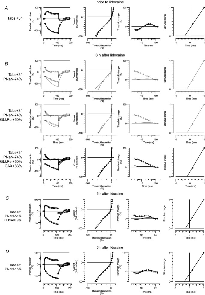
The local anaesthetic lidocaine is known to block voltage-gated Na(+) channels (VGSCs), although at high concentration it was also reported to block other ion channel currents as well as to alter lipid membranes. The aim of this study was to investigate whether the clinical regional anaesthetic action of lidocaine could be accounted for solely by the block of VGSCs or whether other mechanisms are also relevant. We tested the recovery of motor axon conduction and multiple measures of excitability by 'threshold-tracking' after ultrasound-guided distal median nerve regional anaesthesia in 13 healthy volunteers. Lidocaine caused rapid complete motor axon conduction block localized at the wrist. Within 3 h, the force of the abductor pollicis brevis muscle and median motor nerve conduction studies returned to normal. In contrast, the excitability of the motor axons at the wrist remained markedly impaired as indicated by a 7-fold shift of the stimulus-response curves to higher currents with partial recovery by 6 h and full recovery by 24 h. The strength-duration properties were abnormal with markedly increased rheobase and reduced strength-duration time constant. The changes in threshold during electrotonus, especially during depolarization, were markedly reduced. The recovery cycle showed increased refractoriness and reduced superexcitability. The excitability changes were only partly similar to those previously observed after poisoning with the VGSC blocker tetrodotoxin. Assuming an unaltered ion-channel gating, modelling indicated that, apart from up to a 4-fold reduction in the number of functioning VGSCs, lidocaine also caused a decrease of passive membrane resistance and an increase of capacitance. Our data suggest that the lidocaine effects, even at clinical 'sub-blocking' concentrations, could reflect, at least in part, a reversible structural impairment of the axolemma.


Figures

Figure 1
Figure 1. Experimental setup
A, photograph of the experimental setup. Compound muscle action potentials (CMAP) were recorded from the abductor pollicis brevis (APB), the abductor digiti quinti (ADQ) and the flexor digitorum (FD) muscles. The median nerve was stimulated at the wrist and elbow whereas the ulnar nerve was stimulated only at the elbow (proximal to the medial epicondyle). B, diagram of the experimental setup indicating the ulnar nerve anaesthesia followed by the median nerve anaesthesia. C, ultrasound image during lidocaine injection (arrow) near the median nerve. The spread of lidocaine (arrow) over several centimetres along the nerve is depicted in the three-dimensional reconstruction to the right.
Figure 2
Figure 2. Recovery of CMAPs during the first 2 h after lidocaine injection at the median nerve
A, abductor pollicis brevis (APB) CMAPs evoked after stimulation of the median nerve at the wrist (Wr) and elbow (Elb) and flexor digitorum (FD) muscles CMAPs evoked after stimulation of the median nerve at Elb. B and C, the mean time course of recovery of maximal CMAP amplitude (B) and shortest latency (C) was averaged in three subjects in whom the maximal CMAP recovery was studied in details. Error bars represent SEM.
Figure 3
Figure 3. Recovery of motor response threshold from 2 h (2H) to 24 h (24H) after lidocaine as compared to measures prior to lidocaine (PRE)
A, stimulus–response relationships from the wrist (filled circles) and elbow (Elb, triangles) to APB are presented for a single subject before lidocaine injection and at 2 h. Note that in spite of the large right shift in threshold at the wrist, measurements from the elbow remained unchanged. B, mean stimulus–response relationships at the wrist are shown for all subjects at the different time points before and after lidocaine injection. The stimulus current axis is presented on a log scale to facilitate the display of the large changes in threshold. Error bars represent SEM. C–F, dot plots presenting the changes over time of the peak CMAP amplitude (C), threshold latency (D), threshold stimulus (E) and rheobase (F). Hourly means are indicated.
Figure 4
Figure 4. Recovery of motor axon excitability measures after lidocaine block
A–D, mean changes in threshold electrotonus (A), current–threshold relationship (B), recovery cycle (C) and charge–duration relationship (D). Error bars represent SEM. Measurements are presented prior to lidocaine (PRE, grey line), and at 2 h (2H, open circles), 4 h (4H, open triangles) and 24 h (24H, filled circles). E–H, dot plots presenting the changes over time after lidocaine injection of the corresponding measurements in strength–duration time constant, SDTC (E), threshold reduction during depolarizing threshold electrotonus, TEd(90–100 ms) (F), superexcitability of the recovery cycle at 5 ms (G) and threshold reduction during hyperpolarizing threshold electrotonus, TEh(20–40 ms) (H). Hourly means are indicated.
Figure 5
Figure 5. Interpretation of lidocaine effects using the ‘Bostock’ mathematical model
The four corresponding excitability measures are presented in rows from left to right: threshold electrotonus, current–threshold relationship, recovery cycle and charge–duration relationship. Open circles indicate mean excitability measurements recorded prior to lidocaine (A) and at 3 h after lidocaine (B), 5 h after lidocaine (C) and 6 h after lidocaine (D). Note that the first two rows in B are shown in grey as they reflect unsatisfactory intermediate fits. The modelled rheobase is scaled down to the single axon by setting the charge for a 1 ms pulse as 1. The continuous line indicates the modelled excitability measures. The changed parameters from the reference model (Howells et al. 2012) are indicated to the left: absolute temperature (Tabs), VGSC permeability (PNaN), relative leak conductance (GLkRel) and axonal capacitance (CAX).
Figure 6
Figure 6. Effect of lidocaine vehicle
The effect of lidocaine vehicle (Veh.) was compared to the effect of lidocaine (Lid. + Veh.) in two subjects re-examined for this control experiment. A, mean excitability measures (charge–duration (upper left), recovery cycle (upper right), threshold electrotonus (lower left) and the current–threshold relationships (lower right)) were measured before injection (PRE, full lines), immediately after injection (0H, open circles) and at 3 h (3H, filled circles). The vehicle measurements are shown in grey. Note that the measurements at immediately after injection could only be obtained for the vehicle injection. B, photograph showing four consecutive anatomical levels of injection from distal to proximal from the stimulation site. C, corresponding ultrasound scans (approx. 2 cm in width/1.5 cm in height) for one subject. White arrows indicate the fluid volume around the median nerve just after injection.

Similar articles

Cited by

References

    1. Argoff CE. Topical analgesics in the management of acute and chronic pain. Mayo Clin Proc. 2013;88:195–205. - PubMed
    1. Barrett EF, Barrett JN. Intracellular recording from vertebrate myelinated axons: mechanism of the depolarizing afterpotential. J Physiol. 1982;323:117–144. - PMC - PubMed
    1. Bostock H. The strength–duration relationship for excitation of myelinated nerve: computed dependence on membrane parameters. J Physiol. 1983;341:59–74. - PMC - PubMed
    1. Bostock H, Baker M, Reid G. Changes in excitability of human motor axons underlying post-ischaemic fasciculations: evidence for two stable states. J Physiol. 1991;441:537–557. - PMC - PubMed
    1. Bostock H, Cikurel K, Burke D. Threshold tracking techniques in the study of human peripheral nerve. Muscle Nerve. 1998;21:137–158. - PubMed

Publication types

LinkOut - more resources