. 2014 Apr 7;9(4):e93908.
doi: 10.1371/journal.pone.0093908. eCollection 2014.

Gap-directed translesion DNA synthesis of an abasic site on circular DNA templates by a human replication complex

Affiliations

Gap-directed translesion DNA synthesis of an abasic site on circular DNA templates by a human replication complex

Giuseppe Villani et al. PLoS One. .

Abstract

DNA polymerase ε (pol ε) is believed to be the leading strand replicase in eukaryotes whereas pols λ and β are thought to be mainly involved in re-synthesis steps of DNA repair. DNA elongation by the human pol ε is halted by an abasic site (apurinic/apyrimidinic (AP) site). We have previously reported that human pols λ, β and η can perform translesion synthesis (TLS) of an AP site in the presence of pol ε. In the case of pol λ and β, this TLS requires the presence of a gap downstream from the product synthetized by the ε replicase. However, since these studies were conducted exclusively with a linear DNA template, we decided to test whether the structure of the template could influence the capacity of the pols ε, λ, β and η to perform TLS of an AP site. Therefore, we have investigated the replication of damaged "minicircle" DNA templates. In addition, replication of circular DNA requires, beyond DNA pols, the processivity clamp PCNA, the clamp loader replication factor C (RFC), and the accessory proteins replication protein A (RPA). Finally we have compared the capacity of unmodified versus monoubiquitinated PCNA in sustaining TLS by pols λ and η on a circular template. Our results indicate that in vitro gap-directed TLS synthesis by pols λ and β in the presence of pol ε, RPA and PCNA is unaffected by the structure of the DNA template. Moreover, monoubiquitination of PCNA does not affect TLS by pol λ while it appears to slightly stimulate TLS by pol η.


Conflict of interest statement

Competing Interests: The authors have declared that no competing interests exist.

Figures

Figure 1
Figure 1. DNA templates –primers used in this study.
AP indicates the position of the synthetic abasic site at the G position of the undamaged template. For details see Materials and Methods
Figure 2
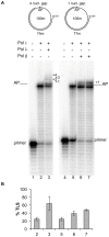
Figure 2. Translesion synthesis over an AP site by DNA Polymerases λ and β in the presence of DNA DNA polymerase ε on circular DNA templates with gaps of four or one nucleotides downstream the lesion.
Experiments were performed with damaged templates with either 74 or 77 mer primers, as depicted on the top of Fig 2A and in Fig 1. The enzymes are indicated at the top of the Figure. Experiments were carried out as described in Materials and Methods. A: Lane 1, no polymerase present. Lane 2, reaction incubated for 35 minutes with 0.025 pmol of pol ε. Lane 3, reaction incubated for 30 minutes with 0.025 pmol of pol ε, then 0.25 pmol of pol λ was added and the incubation was continued for 5 minutes. Lane 4, no polymerase present. Lane 5, reaction incubated for 35 minutes with 0.025 pmol of pol ε. Lane 6, reaction incubated for 30 minutes with 0.025 pmol of pol ε, then 0.25 pmol of pol β was added and the incubation continued for 5 minutes. Lane 7, reaction incubated for 30 minutes with 0.025 pmol of pol ε, then 0.25 pmol of pol β was added and the incubation continued for 10 minutes. The position of the primers, the AP site and the nucleotides past the AP site are indicated. B: Quantification of the percentage of TLS, calculated as described in Materials and Methods. Mean +/− S.D. values for three independent experiments are indicated.
Figure 3
Figure 3. RFC stimulation of PCNA dependent synthesis on a circular DNA template.
Experiments were performed with the intact template and a 44Fig 1. The proteins are indicated at the top of the Figure. Experiments were carried out as described in Materials and Methods. A: Lane 1, reaction incubated for 5 minutes with 0.03 pmol of pol δ and 1 pmol of PCNA. Lane 2, reaction incubated for 5 minutes with 0.03 pmol of pol δ, 1 pmol of PCNA and 80 ngs of RFC. Lane 3, reaction incubated for 5 minutes with 0.03 pmol of pol δ, 1 pmol of PCNA and 160 ngs of RFC. Lane 4, reaction incubated for 5 minutes with 0.03 pmol of pol δ, 1 pmol of PCNA and 240 ngs of RFC. Lane 5, 160 ngs of RFC alone incubated for 25 minutes. Lane 6–10, reactions incubated with 0.03 pmol of pol δ and 1 pmol of PCNA for 5,10,15,20 and 25 minutes respectively. Lane 11–15, reactions incubated with 0.03 pmol of pol δ, 1 pmol of PCNA and 160 ngs of RFC for 5,10,15,20 and 25 minutes respectively. B: Quantification of the data in A by plotting the relative intensity of the bands migrating higher than the primer. The positions of the primer and of the 100 mer full length product are indicated. Left graph shows mean +/− S.D. values for three independent experiments. Right graph shows the quantification of the right panel of figure 3A.
Figure 4
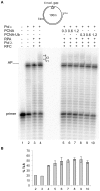
Figure 4. Translesion synthesis over an AP site on circular DNA templates by a reconstituted replication complex.
Experiments were performed with damaged templates primed with either 74 or 77 mer primers, as depicted on the top of Fig 4A and in Fig 1. The proteins are indicated at the top of the Figure. Experiments were carried out as described in Materials and Methods. A: Lane 1, no proteins present. Lane 2, reaction incubated for 35 minutes with 0.025 pmol of pol ε, 1 pmol of PCNA and 0.25 pmol of RPA. Lane 3, reaction incubated for 35 minutes with 0.025 pmol of pol ε, 1 pmol of PCNA, 0.25 pmol of RPA and 160 ngs of RFC. Lane 4, reaction incubated 5 minutes with 0.25 pmol of pol λ, 1 pmol PCNA, 0.25 pmol RPA and 160 ngs of RFC. Lane 5, reaction incubated for 30 minutes with 0.025 pmol of pol ε, 1 pmol of PCNA and 0.25 pmol of RPA; then 0.25 pmol of pol λ was added and the incubation was continued for 5 minutes. Lane 6, reaction incubated for 30 minutes with 0.025 pmol of pol ε, 1 pmol of PCNA, 0.25 pmol of RPA and 160 ngs of RFC; then 0.25 pmol of pol λ was added and the incubation was continued for 5 minutes. Lane 7, no proteins present. Lane 8, reaction incubated for 35 minutes with 0.025 pmol of pol ε, 1 pmol of PCNA and 0.25 pmol of RPA. Lane 9, reaction incubated for 35 minutes with 0.025 pmol of pol ε, 1 pmol of PCNA, 0.25 pmol of RPA and 160 ngs of RFC. Lane 10, reaction incubated 5 minutes with 0.25 pmol of pol β, 1 pmol of PCNA, 0.25 pmol of RPA and 160 ngs RFC. Lane11, reaction incubated for 30 minutes with 0.025 pmol of pol ε, 1 pmol of PCNA and 0.25 pmol of RPA; then 0.25 pmol of pol β was added and the incubation was continued for 5 minutes. Lane 12, reaction incubated for 30 minutes with 0.025 pmol of pol ε, 1 pmol of PCNA, 0.25 pmol of RPA and 160 ngs of RFC; then 0.25 pmol of pol β was added and the incubation was continued for 5 minutes. The position of the primers, the AP site and the nucleotides past the AP site are indicated. B: quantification of the percentage of TLS, calculated as described under Materials and Methods. Mean +/− S.D. values for three independent experiments are indicated.
Figure 5
Figure 5. Monoubiquitination of PCNA does not change the pattern of TLS by DNA polymerase λ.
Experiments were performed with a damaged template primed with the 74 mer primer, as depicted on the top of the Fig 5A and in Fig 1. The proteins are indicated at the top of the figure. Experiments were carried out as described in Materials and Methods. A: lane 1, no proteins present. Lane 2, reaction incubated for 35 minutes with 0.025 pmol of pol ε. Lane 3, reaction incubated for 35 minutes with 0.025 pmol of pol ε, 0.25 pmol of RPA and 160 ngs of RFC. Lane 4, reaction incubated for 30 minutes with 0.025 pmol of pol ε, 0.25 pmol of RPA and 160 ngs of RFC; then 0.25 pmol of pol λ was added and the incubation continued for 5 minutes. Lanes 5 to 7; as in lane 4 but with 0.3, 0.6 and 1.2 pmol of PCNA respectively. Lanes 8 to 10; as in lane 4 but with 0.3, 0.6 and 1.2 pmol of monoubiquitinated PCNA respectively. The position of the primers, the AP site and the nucleotides past the AP site are indicated. B: Quantification of the percentage of TLS, calculated as described in Materials and Methods. Mean +/− S.D. values for three independent experiments are indicated.
Figure 6
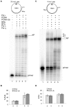
Figure 6. Monoubiquitinated PCNA stimulates TLS by DNA polymerase η.
Experiments were performed with damaged templates primed with the 44 or 74 mer primers, as depicted on the top of Fig 6A and in Fig 1. The proteins are indicated at the top of the Fig. Experiments were carried out as described in Materials and Methods. A: 1, no proteins present. Lane2, reaction incubated for 30 minutes with 0.025 pmol of pol ε, 1.2 pmol of PCNA, 0.25 pmol of RPA and 160 ngs of RFC; then 0.25 pmol of pol λ was added and the incubation continued for 5 minutes. Lane 3, as in lane 2 but with 1.2 pmol of monoubiquinated PCNA. Lane 4, reaction incubated for 30 minutes with 0.025 pmol of pol ε, 1.2 pmol of PCNA, 0.25 pmol of RPA and 160 ngs of RFC; then 0.25 pmol of pol η was added and the incubation continued for 5 minutes. Lane 5, as in lane 4 but with 1.2 pmol of monoubiquinated PCNA. The positions of the primer and of the AP site are indicated. B: quantification of the percentage of TLS of the data shown in A, calculated as described in Materials and Methods. Error bars reflect ranges from two independent experiments. C: 1, no proteins present. Lane2, reaction incubated for 30 minutes with 0.025 pmol of pol ε, 1.2 pmol of PCNA, 0.25 pmol of RPA and 160 ngs of RFC; then 0.25 pmol of pol λ was added and the incubation was continued for 5 minutes. Lane 3, as in lane 2 but with 1.2 pmol of monoubiquinated PCNA. Lane 4, reaction incubated for 30 minutes with 0.025 pmol of pol ε, 1.2 pmol of PCNA, 0.25 pmol of RPA and 160 ngs of RFC; then 0.25 pmol of pol η was added and the incubation was continued for 5 minutes. Lane 5, as in lane 4 but with 1.2 pmol of monoubiquinated PCNA. The positions of the primer and of the AP site are indicated. D: quantification of the percentage of TLS of the data shown in C, calculated as described in Materials and Methods. Error bars reflect ranges from two independent experiments.

Similar articles

Cited by

References

    1. Kornberg A, Baker TA (2005) DNA Replication. 2nd ed. University Science Books.
    1. Hübscher U, Spadari S, Villani G, Maga G (2010) DNA Polymerases: Discovery, Characterization and Functions in Cellular DNA Transactions. 1st ed. World Scientific Publishing Company.
    1. Kunkel TA, Burgers PM (2008) Dividing the workload at a eukaryotic replication fork. Trends Cell Biol 18: 521–527. - PMC - PubMed
    1. Lindahl T (1993) Instability and decay of the primary structure of DNA. Nature 362: 709–715. - PubMed
    1. Friedberg EC, Walker GC, Siede W, Wood RD, Schultz RA, et al... (2005) DNA Repair And Mutagenesis. 2nd ed. ASM Press.

Publication types