. 2011 Jan 14;2(1):107-30.
doi: 10.3390/genes2010107.

Prospects and limitations of using endogenous neural stem cells for brain regeneration

Affiliations

Prospects and limitations of using endogenous neural stem cells for brain regeneration

Naoko Kaneko et al. Genes (Basel). .

Abstract

Neural stem cells (NSCs) are capable of producing a variety of neural cell types, and are indispensable for the development of the mammalian brain. NSCs can be induced in vitro from pluripotent stem cells, including embryonic stem cells and induced-pluripotent stem cells. Although the transplantation of these exogenous NSCs is a potential strategy for improving presently untreatable neurological conditions, there are several obstacles to its implementation, including tumorigenic, immunological, and ethical problems. Recent studies have revealed that NSCs also reside in the adult brain. The endogenous NSCs are activated in response to disease or trauma, and produce new neurons and glia, suggesting they have the potential to regenerate damaged brain tissue while avoiding the above-mentioned problems. Here we present an overview of the possibility and limitations of using endogenous NSCs in regenerative medicine.


Figures

Figure 1
Figure 1
Therapeutic strategies using exogenous and endogenous neural stem cells (NSCs). Schematic drawing of a model for the therapeutic use of exogenous (left) and endogenous (right) NSCs. Exogenous NSCs derived from pluripotent stem cells including embryonic stem cells (ESCs) and induced-pluripotent stem cells (iPSCs) are transplanted into the damaged brain, and differentiate into mature neurons to replace damaged ones. Endogenous NSCs that reside in the subventricular zone of the adult brain continuously generate neurons, which migrate into the damaged area, where they replace damaged neurons.
Figure 2
Figure 2
NSCs in the hippocampus. (a) Location, structure, and neuronal circuitry of the dentate gyrus in the hippocampus of the adult rodent brain. The input to the hippocampus is mainly provided by the entorhinal cortex through the perforant path (gray) to the granule cells (pink) in the dentate gyrus; (b) Neurogenesis in the dentate gyrus. NSCs (blue) and neuronal progenitor cells (light green) reside in the SGZ, where they proliferate, and generate immature new neurons (red) (left). The new neurons migrate into the granule cell layer (middle), where some of them differentiate into mature granule cells (pink), and the rest are eliminated by apoptotic cell death (gray) (right).
Figure 3
Figure 3
NSCs in the SVZ. (a) Location and structure of the SVZ. The SVZ is located at the lateral wall of the lateral ventricle and consists of four types of cells: ependymal cells (purple), which have multiple motile cilia that lie over the surface of the SVZ; multipotent and self-renewable NSCs, which have an astrocytic morphology (blue); neuronal progenitor cells (light green); and migratory new neurons (red). NSCs have an apical membrane that makes direct contact with the ventricle, and extends its processes onto the blood vessels (orange); (b) Generation of new neurons in the SVZ. NSCs (blue) slowly and continuously proliferate to generate new NSCs (self-renewal) and neuronal progenitor cells called “transit-amplifying cells” (light green). The transit amplifying cells proliferate quickly and produce immature new neurons (red); (c) Rapid and long-range migration of new neurons. Newly generated new neurons (red) in the rodent SVZ migrate rapidly and reach the olfactory bulb within a week, where they differentiate into mature interneurons. In the migratory path, called the rostral migratory stream (RMS), the new neurons form an elongated chain-like cluster, and move inside a tunnel formed by astrocytic processes (blue), which sometimes occur along a blood vessel (orange); (d) Chains of new neurons along blood vessels in the RMS. Sagittal brain sections were immunostained with the new neuron marker Dcx (red) and endothelial marker CD31 (light blue). Some of the chains of new neurons were extended along and closely associated with blood vessels; (e) Chains of new neurons surrounded by astrocytic processes in the RMS. Sagittal brain sections were immunostained with Dcx (red) and the astrocytic marker GFAP (light blue). GFAP+ astrocytic processes tightly enclosed the chains of new neurons.
Figure 4
Figure 4
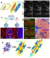
Interaction between migrating new neurons and the surrounding astrocytes. (modified from Kaneko et al., Neuron, 2010 [76]). (a) Expression patterns of Slit1 and Robos in the RMS; (b) Disrupted migration of new neurons in a Slit1-defficient (Slit1−/−) brain slice. Representative paths of new neurons migrating in cultured brain slices obtained by time-lapse imaging were drawn with colored dots and lines. Compared with those of the wild-type (top) slice, the migration of new neurons in the Slit1−/− RMS was irregular and slower (bottom, b'). Scale bars: 200 μm; (c) Slit repels SVZ/RMS astrocytes. Purified astrocytes dissociated from the SVZ and RMS were co-cultured with either Slit-expressing or control HEK cells mixed into collagen gel pieces (top). After 4 days of co-culture, significantly fewer astrocytes stained with GFAP (red) were observed on the Slit-containing gel (bottom right) compared with the control gel (bottom left). Scale bars: 200 μm; (d) Disruption of astrocytic tunnels in the Slit1−/− RMS. Sagittal brain sections containing wild-type or Slit1−/− RMS were immunostained with Dcx and GFAP (left). In the wild-type RMS, GFAP+ (green) astrocytic processes were extended along the chains of new neurons (red), whereas in the Slit1−/− RMS, the arrangement of astrocytic processes was irregular. Electron micrographs (right) reveal an abnormal distribution of astrocytic processes (arrowheads) among the chain-forming new neurons in the Slit1−/− RMS. Scale bars: 50 μm (white); 5 μm (black); (e) Schematic drawings of the Slit-Robo-mediated interaction between new neurons and tunnel-forming astrocytes. The new neuron-secreted Slit1 and astrocyte-expressed Robo receptor control the distribution and arrangement of astrocytes to maintain the migratory path of new neurons, which assists their rapid migration through the RMS.
Figure 5
Figure 5
Migration of new neurons to an injured area. (a) Schematic drawings of SVZ new neurons migrating toward an infarcted area; (b) Mouse brain section 18 days after experimental ischemic stroke stained with the new neuron marker Dcx (brown). Transient middle cerebral artery occlusion (MCAO) caused infarction (white broken line). Eighteen days later, new neurons generated in the SVZ migrated toward the infarcted area. b' shows a higher-magnification image of the boxed area in b; (c) Association of migrating new neurons with the vasculature. A brain section 18 days after MCAO was immunostained with Dcx (green) and the endothelial marker CD31 (red). Many of the new neurons migrating toward the infarcted area were closely associated with blood vessels; (d) Vascular scaffold for new neurons migrating toward the infarcted area. Time-lapse imaging of a cultured brain slice after MCAO. New neurons were labeled by lentivirus injection into the lateral ventricle of Flk1-EGFP mice. A new neuron (green) extended leading process (arrows) and migrated along a blood vessel (red).
Figure 6
Figure 6
Endogenous NSC-derived neuronal regeneration. (a) Schematic drawing of the experimental procedure. The pxCANCre plasmid was injected into the lateral ventricle 5 days before MCAO, then the fate of GFP-labeled new neurons generated in the SVZ was detected 90 days after MCAO; (b) Confocal 3D reconstruction image of a GFP (green)-labeled cell expressing the mature neuronal marker, NeuN (red). Ninety days after MCAO, 29% of the SVZ-derived GFP-positive cells around the infarcted area expressed NeuN, a specific marker for mature neurons. Scale bar: 20 μm; (c) A GFP-positive cell exhibiting a neuronal morphology. Scale bar: 20 μm; (d) An electron micrograph showing a GFP-positive axon (asterisk) containing presynaptic vesicles. A higher-magnification view of the region of the boxed area (d') shows the postsynaptic density (arrowheads). Scale bar: 0.5 μm.

Similar articles

Cited by

References

    1. Gerrard L., Rodgers L., Cui W. Differentiation of human embryonic stem cells to neural lineages in adherent culture by blocking bone morphogenetic protein signaling. Stem Cells. 2005;23:1234–1241. - PubMed
    1. Okada Y., Matsumoto A., Shimazaki T., Enoki R., Koizumi A., Ishii S., Itoyama Y., Sobue G., Okano H. Spatiotemporal recapitulation of central nervous system development by murine embryonic stem cell-derived neural stem/progenitor cells. Stem Cells. 2008;26:3086–3098. - PubMed
    1. Takahashi K., Yamanaka S. Induction of pluripotent stem cells from mouse embryonic and adult fibroblast cultures by defined factors. Cell. 2006;126(4):663–676. - PubMed
    1. Yamanaka S. Strategies and new developments in the generation of patient-specific pluripotent stem cells. Cell Stem Cell. 2007;1(1):39–49. - PubMed
    1. Zhang S.C., Wernig M., Duncan I.D., Brustle O., Thomson J.A. In vitro differentiation of transplantable neural precursors from human embryonic stem cells. Nat. Biotechnol. 2001;19(12):1129–1133. - PubMed

LinkOut - more resources