. 2014 Apr 7;9(4):e92513.
doi: 10.1371/journal.pone.0092513. eCollection 2014.

Computational design of binding proteins to EGFR domain II

Affiliations

Computational design of binding proteins to EGFR domain II

Yoon Sup Choi et al. PLoS One. .

Abstract

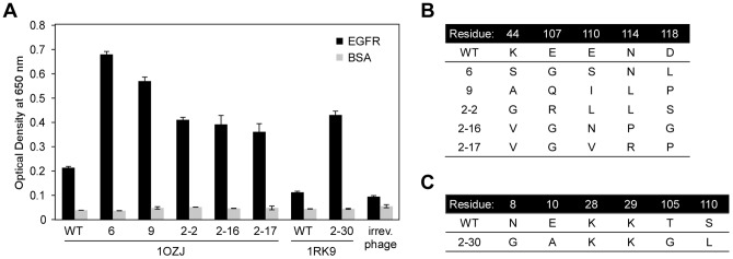
We developed a process to produce novel interactions between two previously unrelated proteins. This process selects protein scaffolds and designs protein interfaces that bind to a surface patch of interest on a target protein. Scaffolds with shapes complementary to the target surface patch were screened using an exhaustive computational search of the human proteome and optimized by directed evolution using phage display. This method was applied to successfully design scaffolds that bind to epidermal growth factor receptor (EGFR) domain II, the interface of EGFR dimerization, with high reactivity toward the target surface patch of EGFR domain II. One potential application of these tailor-made protein interactions is the development of therapeutic agents against specific protein targets.


Conflict of interest statement

Competing Interests: The authors have declared that no competing interests exist.

Figures

Figure 1
Figure 1. Design scheme of target-specific scaffolds.
(A) Synthetic antibodies can achieve extremely diverse structures through sequence randomization of the complementarity determining region (CDR). Among diverse structures, only antibodies with complementary shapes are able to recognize and bind to a particular epitope. (B) By imitating synthetic antibody generation, we devised a strategy to select target-specific scaffolds from the human proteome with shapes that are complementary to the target surface patch. (C) The flow chart shows a two-step strategy to obtain target-specific scaffolds (middle). In the first step, a virtual screening of a human protein scaffold library is conducted to determine a framework specific to the surface patch of interest. Target specific-scaffolds with shapes complementary to the surface patch of interest are selected from the scaffold library through protein docking simulations (upper right). The scaffold–target docking structures with the most favorable complex formation energies are further evaluated (left). In the second step, the scaffold interface in the selected scaffold–target model is optimized by sequence randomization and phage display using directed evolution (lower right).
Figure 2
Figure 2. Scaffolds 1OZJ and 1RK9 have shapes complementary to EGFR domain II.
(A) The inactivated EGFR monomer exists in equilibrium between the tethered and untethered conformations. The binding of EGF (pink) stabilizes the untethered monomers, which exposes the dimerization arm of domain II (green) and activates EGFR to form homodimers at the domain II dimeric interface (domain I, blue; domain II, green; domain III, yellow; domain IV, gray). For clarity, domain II on the right-hand EGFR in the homodimer is shown in orange. (B) EGFR activation can be blocked by binding domain II, which is exposed in the untethered conformation, with the designed scaffold (magenta), which sterically interferes with EGFR dimerization. (C) The docking conformation of 1OZJ–EGFR, which was selected from the virtual screening procedure (left), and the enlarged 1OZJ structure (magenta). (D) The docking conformation of 1RK9–EGFR, which was selected from the virtual screening procedure (left), and the enlarged 1RK9 structure (magenta). Energetically unfavorable residues that were selected for optimization are depicted in blue. (E–F) Amino acid sequences for 1OZJ and 1RK9, respectively. The blue residues represent amino acid residues that were energetically unfavorable for EGFR complex formation. (G–H) The complex formation energy of the scaffold interface residues upon the formation of the 1OZJ–EGFR and 1RK9–EGFR docking complexes, respectively. The blue bars depict the energetically unfavorable residues that were selected for optimization. The horizontal axis shows each amino acid in the scaffold interface, and the vertical axis represents the energy contribution (delta G) to the complex formation.
Figure 3
Figure 3. Strong binders were enriched by directed evolution of the 1OZJ and 1RK9 scaffolds.
(A) The reactivity of the protein binders (wild-type and mutant clones) against EGFR (black) and BSA (gray) as assessed by phage-ELISA. Error bars represent the standard deviation of triplicate measures. (B) The sequence of the mutant clones generated from 1OZJ. (C) The sequence of the mutant clone generated from 1RK9.
Figure 4
Figure 4. Binders generated from the 1OZJ and 1RK9 scaffolds bind to EGFR fragments I–IV and I–II.
The reactivity of the protein binders (wild-type and mutant clones) against EGFR domain I–IV (dark blue) and EGFR domain I–II (light blue) as assessed by phage-ELISA. Error bars represent the standard deviation of triplicate phage-ELISA experiments.
Figure 5
Figure 5. Reactivity of the binders to EGFR depended on the presence of EGFR domain II.
(A) Chimeric EGFR fragments were generated with the EGFR domain II replaced with domain II of either ErbB2 (orange) or ErbB4 (dark blue). (B) The reactivity of the protein binders to the EGFR fragments containing EGFR domain II (green), the chimeric EGFR fragments with ErbB2 domain II (orange), and the chimeric EGFR fragments with ErbB4 domain II (dark blue), as assessed by phage-ELISA. Error bars represent the standard deviation of triplicate phage-ELISA experiments.
Figure 6
Figure 6. Binders have a higher reactivity to EGFR in the presence of ligand EGF.
(A) Ligand EGF stabilizes the untethered form of EGFR to expose domain II and increase scaffold binding (domain I, blue; domain II, green; domain III, yellow; domain IV, gray; EGF ligand, pink; scaffold, red). (B) The reactivity to EGFR in the presence of EGF (pink) or the absence of EGF (black). (C) The reactivity to EGFR fragment containing domains I–IV in the presence of EGF (orange) or the absence of EGF (black) as assessed by phage-ELISA. Error bars represent the standard deviation of phage-ELISA experiments performed with eight-fold replication.

References

    1. Yamada T, Bork P (2009) Evolution of biomolecular networks: lessons from metabolic and protein interactions. Nat Rev Mol Cell Biol 10: 791–803. - PubMed
    1. Berg J, Lassig M, Wagner A (2004) Structure and evolution of protein interaction networks: a statistical model for link dynamics and gene duplications. BMC Evol Biol 4: 51. - PMC - PubMed
    1. Levy ED, Pereira-Leal JB (2008) Evolution and dynamics of protein interactions and networks. Curr Opin Struct Biol 18: 349–357. - PubMed
    1. Wagner A (2003) How the global structure of protein interaction networks evolves. Proc Biol Sci 270: 457–466. - PMC - PubMed
    1. Kim PM, Lu LJ, Xia Y, Gerstein MB (2006) Relating three-dimensional structures to protein networks provides evolutionary insights. Science 314: 1938–1941. - PubMed

Publication types