. 2014 Jun;34(12):2249-63.
doi: 10.1128/MCB.01413-13. Epub 2014 Apr 7.

Multisite phosphorylation of the Sum1 transcriptional repressor by S-phase kinases controls exit from meiotic prophase in yeast

Affiliations

Multisite phosphorylation of the Sum1 transcriptional repressor by S-phase kinases controls exit from meiotic prophase in yeast

Daniel Corbi et al. Mol Cell Biol. 2014 Jun.

Abstract

Activation of the meiotic transcription factor Ndt80 is a key regulatory transition in the life cycle of Saccharomyces cerevisiae because it triggers exit from pachytene and entry into meiosis. The NDT80 promoter is held inactive by a complex containing the DNA-binding protein Sum1 and the histone deacetylase Hst1. Meiosis-specific phosphorylation of Sum1 by the protein kinases Cdk1, Ime2, and Cdc7 is required for NDT80 expression. Here, we show that the S-phase-promoting cyclin Clb5 activates Cdk1 to phosphorylate most, and perhaps all, of the 11 minimal cyclin-dependent kinase (CDK) phospho-consensus sites (S/T-P) in Sum1. Nine of these sites can individually promote modest levels of meiosis, yet these sites function in a quasiadditive manner to promote substantial levels of meiosis. Two Cdk1 sites and an Ime2 site individually promote high levels of meiosis, likely by preparing Sum1 for phosphorylation by Cdc7. Chromatin immunoprecipitation reveals that the phosphorylation sites are required for removal of Sum1 from the NDT80 promoter. We also find that Sum1, but not its partner protein Hst1, is required to repress NDT80 transcription. Thus, while the phosphorylation of Sum1 may lead to dissociation from DNA by influencing Hst1, it is the presence of Sum1 on DNA that determines whether NDT80 will be expressed.


Figures

FIG 1
FIG 1
Changes to the Sum1 and Sum1-ci proteins during meiotic development. (A) Sum1-HH (wt) and Sum1-ci-HH (ci) proteins were enriched on Ni beads from ndt80Δ cells collected at the indicated times after transfer to sporulation medium, and the proteins were analyzed by electrophoresis and immunoblot analyses using an HA antibody (Sum1). A long and a short exposure of the same blot are shown to facilitate the comparison. The same samples were analyzed with a phospho-specific antiserum for the residue that is phosphorylated by Ime2 (pT306) and a polyhistidine antiserum as a loading control (Snf1). (B) Sum1-HH proteins containing the indicated mutations from vegetative (V) or meiotic (M) ndt80Δ cells were analyzed as in panel A except that Phos-tag acrylamide was included in the running gel. The Sum1 proteins from 4 times as many meiotic as vegetative cells were analyzed to facilitate comparison. (C) Occupancies of wild-type Sum1 and Sum1-ci were analyzed at the SMK1 and NDT80 promoters in vegetative and meiotic ndt80Δ cells using a Sum1 antibody or a negative-control IgG as indicated. The occupancy of Sum1 in vegetative cells was arbitrarily set at 100.
FIG 2
FIG 2
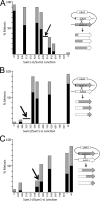
Directional phosphosite analysis (DPA) of Sum1 chimeras. Starting strains in each case consist of duplicated SUM1 alleles flanking a URA3 marker which are diagrammed in a looped configuration (right). The X indicates recombination, which can occur throughout the indicated region of homology to generate the chimeric products indicated below. Recombinants in SUM1 were selected using 5-FOA, and the recombination interval was identified by DNA sequencing (x axis). In each case, the data bar is positioned between the phosphoacceptor residues that were either absent (sum1-ci) or present (SUM1). Recombinants were crossed by a sum1-ci tester strain, and the fraction of cells that completed a single meiotic division (gray; 2 DAPI-stained foci) or more than one division (black; 3 or 4 DAPI-stained foci) was scored (y axis). The arrows point to inflection intervals discussed in the text. (A) Analysis of nonphosphorylatable/phosphorylatable Sum1-ci/Sum1 chimeras. (B) Analysis of phosphorylatable/nonphosphorylatable Sum1/Sum1-ci chimeras. (C) Analysis of phosphorylatable/nonphosphorylatable Sum1-i/Sum1-ci chimeras.
FIG 3
FIG 3
Mutants in SUM1 reveal 2 classes of CDK phospho-consensus sites. (A) Homozygous strains of the indicated genotypes were assayed for meiosis at 24 h postinduction (gray, 2 DAPI-stained foci; black, 3 or 4 DAPI-stained foci; 200 cells counted per experiment, n = 3). The 3A mutant contains phosphosite substitutions of the 3 major phosphoacceptors (T306A, S379A, and S512A). The sum1-ci-306T mutant is identical to sum1-c, as indicated (c). (B) The indicated SUM1 mutants in the ndt80Δ background were transferred to sporulation medium, cells were withdrawn at the indicated times (hours), and Smk1-HA was measured by immunoblot analyses as a surrogate assay for Sum1 removal. The PSTAIR antibody, which recognizes a doublet of Cdk1 and Pho85, was used as a loading control.
FIG 4
FIG 4
The recombination checkpoint does not influence the phosphorylation of Sum1 by Ime2 or Cdk1, and Clb5 is the cyclin that activates Cdk1 for Sum1 phosphorylation. (A) Sum1-HH purified from the indicated strains that were grown vegetatively (V) or incubated in SPO medium for 5.5 h (M) was analyzed by Phos-tag electrophoresis and immunoblot analysis using a Sum1 antiserum or a phospho-specific antiserum for pT306. Sum1 from 4.5 times more meiotic than vegetative cells was analyzed. (B) Cells of the indicated genotype were collected at the indicted times after transfer to sporulation medium and analyzed by immunoblotting for Smk1-HA protein as a surrogate assay for Sum1 downregulation. PSTAIR immunoreactivity was used to control for loading. (C) Strains of the indicated genotypes were assayed for the production of Smk1-HA during vegetative growth (V) and following incubation for 7 h in sporulation medium (M). PSTAIR immunoreactivity was used to control for loading. (D) Strains of the indicated genotype were transferred to sporulation medium, fixed and stained with DAPI at 24 h postinduction, and photographed using phase-contrast microscopy (phase) or fluorescence microscopy (DAPI). Note the appearance of spore-like structures (black arrow) and the diffuse DNA masses (white arrow) in the clb5Δ strain compared to the single DNA masses and the absence of spore-like structures in the clb5Δ sum1-i strain.
FIG 5
FIG 5
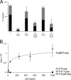
Major phosphoregulatory site function requires −1 S residues. (A) Homozygous sum1 strains containing the indicated substitutions were assayed for meiosis (gray, 2 DAPI-stained foci; black, 3 or 4 DAPI-stained foci; n = 3, 200 cells counted per experiment). (B) The indicated peptides were assayed in phosphotransferase reactions using purified Cdc7/Dbf4 (-S-S-P-pep, the 372–384 Cdk1 phospho-consensus peptide; -S-pS-P-pep, the 372–384 peptide phosphorylated on the Cdk1 site; R-P-S-T-pep, the 299–312 Ime2 phosphoacceptor peptide; R-P-S-pT-pep, the 299–312 peptide phosphorylated on the Ime2 site).
FIG 6
FIG 6
Cdc7 is required for the NDT80-dependent and NDT80-independent pathways for Sum1 removal and shows synthetic interactions with sum1 phosphosite mutants. (A) cdc7-as3-myc SMK1-HA cells were transferred to sporulation medium, inhibitor (PP1) was added at the indicated time, and cells were withdrawn at 7 h postinduction and tested for Smk1-HA production as a surrogate assay for Sum1 repression (N, no PP1 added). Cdc7-as3-Myc was measured using a Myc antibody to control for protein levels. (B) Cells of the indicated SUM1 genotype in the CDC7 wild-type (squares) and cdc7-as3-myc (triangles) backgrounds were transferred to sporulation medium lacking inhibitor, withdrawn at the indicated time postinduction, and assayed for the completion of meiosis (combined MI and MII values). (C) The indicated SUM1 alleles in the cdc7-as3-myc background were treated with PP1 at 3 h postinduction (black bars) or not treated (gray bars), and cells were collected at 24 h and scored for meiosis (MI and MII). In all cases, further increases in meiosis did not take place after the 24-h time point.
FIG 7
FIG 7
NDT80 expression is repressed by Sum1 in an Hst1-independent manner. Cells of the indicated genotype were grown to mid-log phase in rich medium, total RNA was prepared, and the indicated transcript was assayed by Northern blotting analyses. Probes specific for the Ume6-repressible early meiosis-specific gene HOP1, the Sum1-repressible SMK1 gene, and the Ume6/Sum1-coregulated NDT80 gene are shown. The ethidium-stained rRNAs are shown as loading controls. EtBr, ethidium bromide.
FIG 8
FIG 8
Regulatory phosphosites in Sum1. The minimal Cdk1 phospho-consensus S/T-P sites (at residues 242, 313, 315, 318, 379, 409, 512, 616, 697, 738, and 817) that were changed to A-P in sum1-c are indicated with black lines. The Ime2 phosphoacceptor (at residue T306) that was changed to an A residue in sum1-i is shown in orange. Sites in green are potential Cdc7 phosphoacceptors that require prior phosphorylation of adjacent sites by Clb5/Cdk1. The question mark at the 305 site reflects uncertainty about whether this residue is a Cdc7 target. The leftward shaded column indicates Sum1 activity which is inversely related to the probability of prophase exit and completion of meiotic development as indicated by the rightward shaded column. The downward arrows reflect relative potencies of each of the phosphoacceptor sites.

References

    1. Park KS, Mohapatra DP, Misonou H, Trimmer JS. 2006. Graded regulation of the Kv2.1 potassium channel by variable phosphorylation. Science 313:976–979. 10.1126/science.1124254 - DOI - PubMed
    1. Koivomagi M, Valk E, Venta R, Iofik A, Lepiku M, Balog ER, Rubin SM, Morgan DO, Loog M. 2011. Cascades of multisite phosphorylation control Sic1 destruction at the onset of S-phase. Nature 480:128–131. 10.1038/nature10560 - DOI - PMC - PubMed
    1. Nobles KN, Xiao K, Ahn S, Shukla AK, Lam CM, Rajagopal S, Strachan RT, Huang TY, Bressler EA, Hara MR, Shenoy SK, Gygi SP, Lefkowitz RJ. 2011. Distinct phosphorylation sites on the beta(2)-adrenergic receptor establish a barcode that encodes differential functions of beta-arrestin. Sci. Signal. 4:ra51. 10.1126/scisignal.2001707 - DOI - PMC - PubMed
    1. Winter E. 2012. The Sum1/Ndt80 transcriptional switch and commitment to meiosis in Saccharomyces cerevisiae. Microbiol. Mol. Biol. Rev. 76:1–15. 10.1128/MMBR.05010-11 - DOI - PMC - PubMed
    1. Simchen G. 2009. Commitment to meiosis: what determines the mode of division in budding yeast? Bioessays 31:169–177. 10.1002/bies.200800124 - DOI - PubMed

Publication types

MeSH terms

LinkOut - more resources