. 2014 Mar 10:4:4329.
doi: 10.1038/srep04329.

Microglia trigger astrocyte-mediated neuroprotection via purinergic gliotransmission

Affiliations

Microglia trigger astrocyte-mediated neuroprotection via purinergic gliotransmission

Youichi Shinozaki et al. Sci Rep. .

Abstract

Microglia are highly sensitive to even small changes in the brain environment, such as invasion of non-hazardous toxicants or the presymptomatic state of diseases. However, the physiological or pathophysiological consequences of their responses remain unknown. Here, we report that cultured microglia sense low concentrations of the neurotoxicant methylmercury (MeHg(low)) and provide neuroprotection against MeHg, for which astrocytes are also required. When exposed to MeHg(low), microglia exocytosed ATP via p38 MAPK- and vesicular nucleotide transporter (VNUT)-dependent mechanisms. Astrocytes responded to the microglia-derived ATP via P2Y1 receptors and released interleukin-6 (IL-6), thereby protecting neurons against MeHg(low). These neuroprotective actions were also observed in organotypic hippocampal slices from wild-type mice, but not in slices prepared from VNUT knockout or P2Y1 receptor knockout mice. These findings suggest that microglia sense and respond to even non-hazardous toxicants such as MeHg(low) and change their phenotype into a neuroprotective one, for which astrocytic support is required.


Figures

Figure 1
Figure 1. IL-6 release from astrocytes, microglia and mixed glial cultures.
(a) ELISA analysis of MeHg-induced IL-6 release from astrocytes. MeHghigh (1 or 3 μM, 24 h) increased in the amount of IL-6 release from Rt astrocytes but not by MeHglow (0.01 or 0.1 μM, 24 h) (n = 26, **p < 0.01, one-way ANOVA followed by Fisher's LSD test). (b) Microglia released no IL-6 in response to MeHg. At no concentration did Rt microglia monocultures show significant IL-6 release (n = 12, p > 0.05, one-way ANOVA followed by Fisher's LSD test). (c) Rt microglia/astrocyte mixed cultures showed IL-6 release in response to both MeHglow and MeHghigh (0.01–3 μM) (n = 8, *p < 0.05, **p < 0.01 vs. control, one-way ANOVA followed by Fisher's LSD test). For evaluating IL-6 production from glial cell cultures, the cells were serum starved for 24 h before MeHg stimulation. (d) Conditioned media from mixed glial cultures showed a neuroprotective effect on primary cortical neurons against MeHg. Reduced neuronal viability evoked by MeHglow (0.1 μM, 48 h) was restored by MACM (n = 4–8, **p < 0.01 vs. no treatment group; ##p < 0.01 vs. MeHglow, one-way ANOVA followed by Fisher's LSD test). Without MeHg, MACM did not affect basal neuronal viability. Values are the means ± SEM for all groups.
Figure 2
Figure 2. ATP is released from astrocytes, microglia, and mixed glial cultures.
(a) Rt astrocyte monocultures released ATP in response to MeHghigh (1 or 3 μM for 3 h) but not to MeHglow (i.e., 0.01 or 0.1 μM for 3 h) (n = 15–24, **p < 0.01 vs. control, one-way ANOVA followed by Fisher's LSD test). (b) Microglia released ATP in response to a limited concentration window. MeHghigh did not induce ATP release from Rt microglia monocultures. At 0.1 μM, MeHg significantly increased the extracellular ATP level (n = 24–28, **p < 0.01 vs. control, one-way ANOVA followed by Fisher's LSD test). (c) Mixed glial cultures released ATP in response to both MeHglow and MeHghigh. Microglia/astrocyte mixed cultures released ATP in response to 0.1–3 μM of MeHg (for 3 h) (n = 10–16, *p < 0.05, **p < 0.01 vs. control, one-way ANOVA followed by Fisher's LSD test). Glial cells were serum starved for 24 h before MeHg treatment. Values are the means ± SEM for all groups.
Figure 3
Figure 3. MeHglow increases astrocytic Ca2+ oscillations in mixed glial cultures via P2 receptor activation.
(a) Typical Ca2+ responses in microglia/astrocyte mixed cultures in response to MeHglow (0.1 μM, 15 min). Ca2+ images before (control) and after (MeHg) MeHglow are shown. Scale bar: 50 μm. (b) The MeHglow(0.1 μM, 15 min treatment)-evoked increase in the frequencies of Ca2+ oscillations seen in astrocytes in the mixed glial cultures, which was suppressed by the P2 receptor antagonist suramin (100 μM, 30 min pretreatment) (n = 110–240, **p < 0.01 vs. astrocytes/microglia without MeHg, ##p < 0.01 vs. astrocytes/microglia with MeHglow, one-way ANOVA followed by Fisher's LSD test). Astrocytes in the absence of microglia showed no Ca2+ responses to MeHglow (0.1 μM, 15 min). Values are the means ± SEM for (b).
Figure 4
Figure 4. Phosphorylation of p38 MAPK contributes to ATP release from microglia.
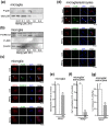
(a) MeHg (2 h treatment) at 0.1 or 0.5 μM evoked phosphorylation of p38 MAPK (P-p38 MAPK) in Rt microglia monocultures. (b) JNK MAPK was not phosphorylated by MeHg. P-ERK1/2 MAPK was rather decreased by MeHg at concentrations over 1.0 μM. The β-actin level was also decreased at concentrations of MeHg over 0.5 μM. Western blot data were cropped and the selected areas were shown here and full-length data are presented in Supplementary Figure 5. The gels have been run under the same experimental conditions. (c) Immunocytochemistry of phosphorylated MAPKs in a Rt microglia monoculture. P-p38 MAPK signal was increased (arrows) after MeHglow treatment (0.1 μM, 2 h). P-ERK1/2 or P-JNK MAPKs did not show enhanced signals in the presence of MeHglow. Scale bar: 10 μm. (d) Phosphorylation of MAPKs in a Rt mixed glial culture. The intensity of P-p38 MAPK signal was increased in microglia (arrows) by MeHglow. P-ERK1/2 MAPK signals were also enhanced but were localized in CD11b-negative cells. The P-JNK MAPK signal intensity was not changed. Scale bar: 20 μm. (e) ATP release is regulated by p38 MAPK. The MeHglow (0.1 μM, 3 h)-induced Rt microglial ATP release was significantly suppressed by a p38 MAPK inhibitor SB203580 (10 μM, 15 min pretreatment) (n = 8, **p < 0.01, Mann–Whitney U-test). (f) The MeHglow-evoked astrocytic Ca2+ oscillation in the Rt glial mixed cultures is regulated by p38 MAPK. The increase in astrocytic Ca2+ oscillations induced by MeHglow (0.1 μM, 15 min) was reduced in the presence of SB203580 (10 μM, 15 min pretreatment) (n = 50, **p < 0.01, Mann–Whitney U-test). (g) IL-6 production from glial mixed cultures is regulated by p38 MAPK. IL-6 production in MeHglow (0.1 μM, 24 h)-treated Rt mixed glial cultures was suppressed by SB203580 (10 μM, 15 min pretreatment) (n = 8, **p < 0.01, Mann–Whitney U-test). Values are the means ± SEM for (e–g). Microglia monocultures and glial mixed cultures were serum starved for 24 h before each experiment.
Figure 5
Figure 5. VNUT-mediated ATP release from microglia is essential for Ca2+ signaling and IL-6 production from astrocytes in the mixed glial cultures.
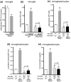
(a) Dependency of of the MeHglow-evoked ATP release on exocytosis. BoNT (5 units/ml, 24 h pretreatment) significantly suppressed the MeHglow-induced ATP release from microglia (n = 5, **p < 0.01, Mann–Whitney U-test). (b) Dependency of the MeHglow-evoked ATP release on VNUT. In contrast to ATP release from WT microglia, VNUTKO microglia showed no ATP release in response to MeHglow (0.1 μM, 3 h) (n = 6–11, **p < 0.01 vs. WT microglia without MeHg, ##p < 0.01 vs. WT microglia with MeHglow, one-way ANOVA followed by Fisher's LSD test). (c) Microglial VNUT is essential for the MeHglow-evoked ATP release in Ms mixed glial cultures. MeHglow(0.1 μM, 3 h) evoked ATP release in WT microglia/WT astrocyte mixed cultures but not in VNUTKO microglia/WT astrocyte cultures (n = 8, **p < 0.01 vs. WT microglia/WT astrocytes without MeHg, ##p < 0.01 vs. WT microglia/WT astrocytes with MeHglow, one-way ANOVA followed by Fisher's LSD test). (d) Microglial VNUT is indispensable for MeHglow-triggered Ca2+ oscillations in astrocytes in the Ms mixed glial cultures. MeHglow (0.1 μM, 15 min) evoked a dramatic increase in the frequency of Ca2+ oscillations in astrocytes in WT microglia/WT astrocyte mixed cultures, but a small increase in VNUTKO microglia/WT astrocytes (n = 98, **p < 0.01 vs. WT microglia/WT astrocytes without MeHg, ##p < 0.01 vs. WT microglia/WT astrocytes with MeHglow, one-way ANOVA followed by Fisher's LSD test). (e) Microglial VNUT-dependent IL-6 production in Ms mixed glial cultures. MeHglow (0.1 μM, 24 h) evoked IL-6 production in WT microglia/WT astrocyte mixed cultures, but not in VNUTKO microglia/WT astrocytes (n = 3, **p < 0.01 vs. WT microglia/WT astrocytes without MeHg, ##p < 0.01 vs. WT microglia/WT astrocytes with MeHglow, one-way ANOVA followed by Fisher's LSD test). Values are the means ± SEM for all groups.
Figure 6
Figure 6. Astrocytic P2Y1 receptors are essential for IL-6 production from mixed glial cultures.
(a) The enhancement of Ca2+ oscillations in astrocytes in the Ms mixed glial culture is mediated by astrocytic P2Y1 receptors. MeHglow (0.1 μM, 15 min treatment) dramatically increased the frequency of astrocytic Ca2+ oscillations in WT microglia/WT astrocyte cultures but not in co-cultures of WT microglia and P2Y1KO astrocytes (n = 98, **p < 0.01 vs. WT microglia/WT astrocytes without MeHg, ##p < 0.01 vs. WT microglia/WT astrocytes with MeHglow, one-way ANOVA followed by Fisher's LSD test). (b) Activation of P2Y1 receptors in astrocytes is essential for IL-6 production in Ms mixed glial cultures. MeHglow (0.1 μM, 24 h) increased IL-6 production in WT microglia/WT astrocytes but not in WT microglia/P2Y1KO astrocytes (n = 3, **p < 0.01 vs. WT microglia/WT astrocytes without MeHg, ##p < 0.01 vs. WT microglia/WT astrocytes with MeHglow, one-way ANOVA followed by Fisher's LSD test). Cells were serum starved for 24 h before MeHg treatment. Values are the means ± SEM for all groups.
Figure 7
Figure 7. ATP-triggered trilateral communication pathways among microglia, astrocytes and neurons in organotypic hippocampal slice cultures.
(a), (b) The endogenous neuroprotection is mediated by glial cells, P2Y1 receptors and IL-6. MeHglow (0.1 μM, 48 h) did not cause an increase in PI incorporation in WT slices. The PI incorporation was increased by minocycline (mino, 10 μM, 24 h pretreatment) or fluorocitrate (FC, 1 μM, 24 h pretreatment), MRS2179 (MRS, 10 μM, 24 h pretreatment) and an IL-6 blocking antibody (IL6Ab, 200 pg/ml, simultaneous treatment) (n = 8–11, **p < 0.01 vs. control). (c) MeHglow (0.1 μM, 24 h) increased IL-6 production from WT slices (n = 4, **p < 0.01 vs. control). (d) MeHglow (0.1 μM, 3 h) increased ATP release from WT slices (n = 5, **p < 0.01 vs. control). (e), (f) MeHglow (0.1 μM, 48 h) increased PI incorporation in VNUTKO slices, which was prevented by ATP (100 μM, 30 min pretreatment) or recombinant IL-6 protein (100 pg/ml, simultaneous treatment) (n = 17–22, **p < 0.01 vs. control, ##p < 0.01 vs. MeHglow). (g) VNUTKO slices did not release ATP in response to MeHglow (0.1 μM, 3 h) (n = 6, p = 0.41). (h) MeHglow (0.1 μM, 24 h) did not induce any increase in IL-6 production from VNUTKO slices (n = 4, p = 0.87). (i), (j) Similar to the findings in VNUTKO slices, MeHglow (0.1 μM, 48 h) increased PI incorporation in P2Y1KO slices; however, unlike the results in VNUTKO slices, exogenously applied ATP (100 μM, 30 min pretreatment) had no effect on MeHglow-increased PI incorporation. Recombinant IL-6 protein (100 pg/ml, simultaneous treatment) significantly reduced the PI signals (n = 8–11, **p < 0.01 vs. control, ##p < 0.01 vs. MeHglow). (k) MeHglow (0.1 μM, 24 h) did not induced IL-6 production from P2Y1KO slices (n = 4, p = 0.19). Values are the mean ± SEM for (b–d), (f–h), (j) and (k). Student's t-test and one-way ANOVA followed by Fisher's LSD test were used for comparisons of two and multiple groups, respectively.
Figure 8
Figure 8. Schematic diagram illustrating the communication pathways among microglia, astrocytes and neurons responsible for neuroprotection.
When exposed to MeHglow, microglia sense MeHglow and activation of p38 MAPK occurs. This is followed by VNUT-dependent ATP exocytosis. The microglia-derived ATP in turn activates P2Y1 receptors and induces Ca2+ oscillations in astrocytes. The activation of P2Y1 receptors induces IL-6 release from astrocytes, thereby leading to neuroprotection against MeHg.

Similar articles

Cited by

References

    1. Tremblay M. E. et al. The role of microglia in the healthy brain. J Neurosci 31, 16064–16069 (2011). - PMC - PubMed
    1. Nimmerjahn A., Kirchhoff F. & Helmchen F. Resting microglial cells are highly dynamic surveillants of brain parenchyma in vivo. Science 308, 1314–1318 (2005). - PubMed
    1. Davalos D. et al. ATP mediates rapid microglial response to local brain injury in vivo. Nat Neurosci 8, 752–758 (2005). - PubMed
    1. Ouchi Y. et al. Microglial activation and dopamine terminal loss in early Parkinson's disease. Ann Neurol 57, 168–175 (2005). - PubMed
    1. Kingwell K. Neurodegenerative disease: Microglia in early disease stages. Nat Rev Neurol 8, 475 (2012). - PubMed

Publication types

MeSH terms

Substances

LinkOut - more resources