

doi: 10.3390/cells1030520.
Autophagy and cancer
Affiliations
- PMID: 24710488
- PMCID: PMC3901115
- DOI: 10.3390/cells1030520
Item in Clipboard
Autophagy and cancer
Cells.
.
Abstract
Autophagy is a housekeeping survival mechanism with a protective function against stress conditions. However, when stress severity or duration increases, it may promote cell death. Paradoxically, autophagy favors cancer development, since cancer cells could enhance their proliferation potential (thus becoming able to resist anticancer therapy) thanks to the energetic supply provided by organelle degradation typically driven by autophagy following a stepwise pathway. The main actors of the autophagic machinery as well as the features shared with apoptosis will be described. Special attention will be paid to the effects of autophagy manipulation.
Figures
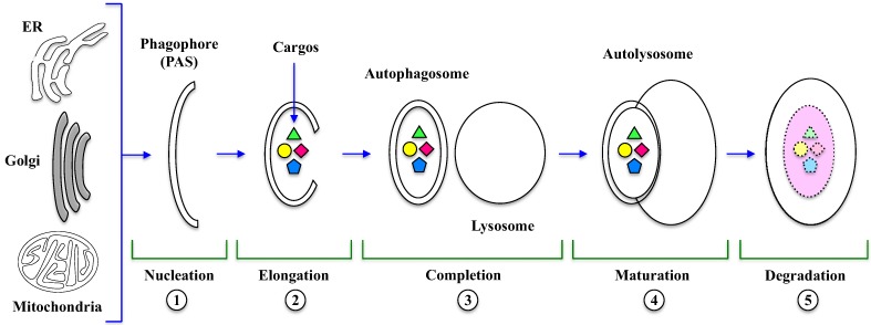
Autophagosome
and
autolysosome
formation. Several morphological changes occur during autophagy, which is stepwise regulated. In the Nucleation (1) and Elongation (2) steps, phagophore originates from membranes of organelles (ER, Golgi, mitochondria) and then encloses the cytosolic cargos, including long-lived, misfolded proteins and damaged organelles, leading to the formation of the autophagosome Completion (3). The Maturation (4) step consists in the fusion of autophagosome with lysosome to form the autolysosome. Finally, during degradation (5), lysosomal hydrolases digest autolysosomal content and release products in the cytosol. PAS: Phagophore Assembly Site; ER: endoplasmic reticulum.
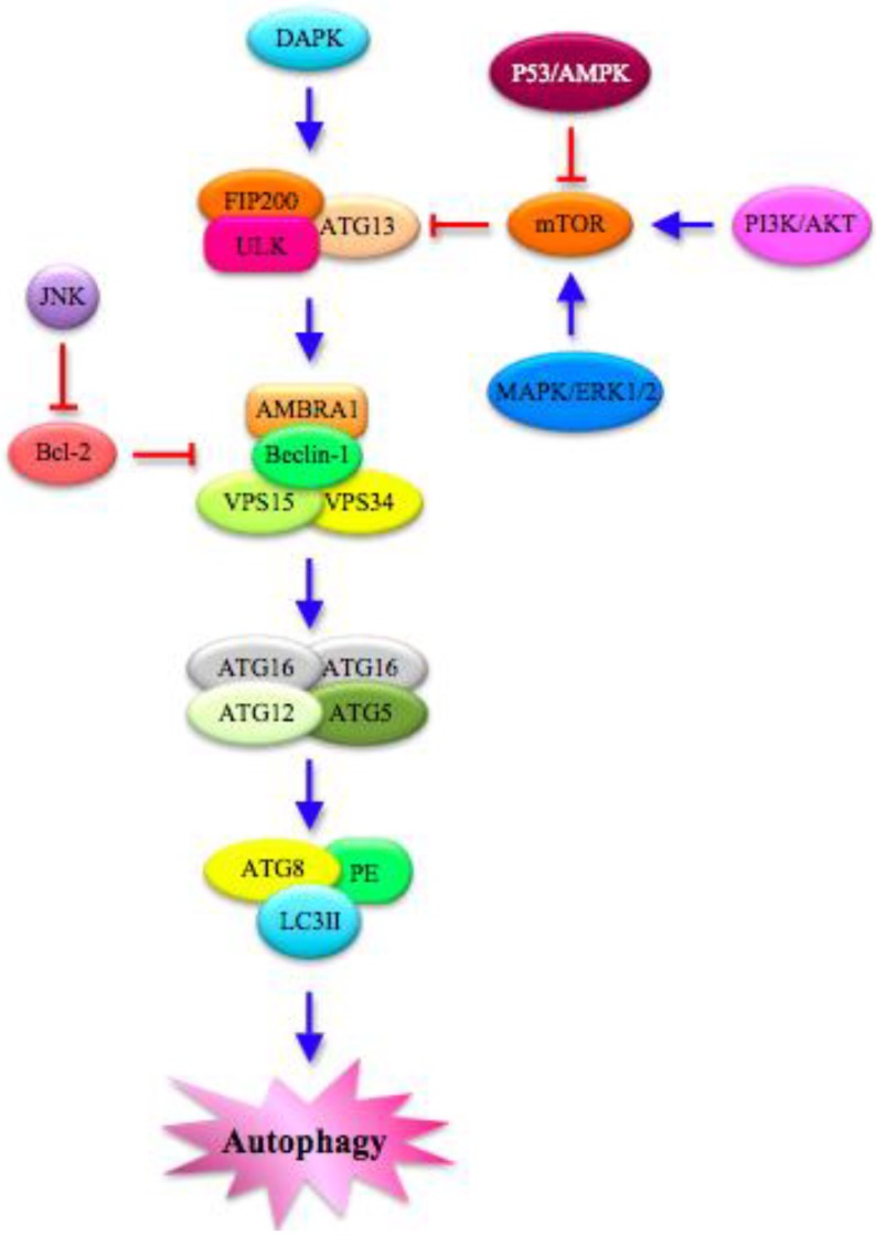
Autophagy
pathway:
Molecular
features. Several proteins in the cytoplasm interact to regulate autophagy. The different steps of the process are modulated by MAPKs (Mitogen-Activated Protein Kinases), such as ERK (Extracellular Signal-Related Kinase) 1/2 and JNK (c-Jun N-terminal Kinase), which mainly act on the key negative regulator of autophagy mTOR (mammalian Target OF Rapamycin). mTOR is also controlled directly by PI3K/AKT (PhosphatidylInositol 3-Kinase/Protein Kinase B) and p53/AMPK (AMP-Activated Protein Kinase) signaling pathways. Moreover, DAPK (Death-Associated Protein Kinase) is implicated in the autophagic cascade, which starts with the formation of ULK (Unc-51-Like Kinase) complex, which is composed of FAK (Focal Adhesion Kinase)-family Interacting Protein of 200 kDa (FIP200), ULK and ATG13. In turn, ULK complex phosphorylates AMBRA1 (Activating Molecule in Beclin-1-Regulated Autophagy), leading to the activation of a complex that includes P-AMBRA1, Beclin-1, VPS (Vacuolar Protein Sorting) 15 and 34. The final steps are characterized by the assembly of ATG complexes made by ATG factors and autophagosome proteins. PE: Phosphatidyl Ethanolamine; LC3II: lipidated form of Microtubule-Associated Protein Light Chain 3.
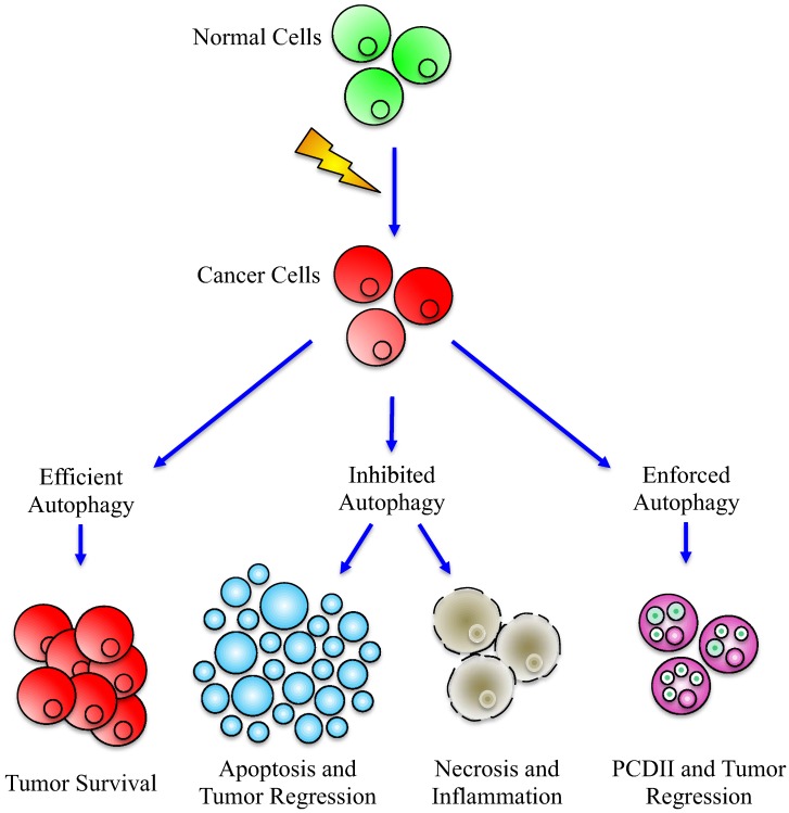
Effects
of
efficient
or
deregulated
Autophagy
in
cancer
development. Alteration of autophagy may have different effects on cancer cells: when efficient, the autophagic machinery could help cancer cells to survive and proliferate (left part), while, when inhibited, autophagy cannot anymore sustain cancer progression, leading to the activation of apoptosis or to necrosis (central part) and, by consequence, tumor regression. Paradoxically, the same end point can be reached after enforced activation of autophagy (right part), which can act as type II PCD (Programmed Cell Death).
References
-
- Ferlay J., Shin H.R., Bray F., Forman D., Mathers C., Parkin D.M. Cancer Incidence and Mortality Worldwide: IARC CancerBase No. 10
-
- Philchenkov A. Apoptosis, cancer, and beyond. Cell Death Differ. 2006;13:2004–2005. doi: 10.1038/sj.cdd.4402009. - DOI
LinkOut - more resources
Full Text Sources
