. 2014 May;109(5):748-56.
doi: 10.1038/ajg.2014.48. Epub 2014 Apr 8.

Novel role of the serine protease inhibitor elafin in gluten-related disorders

Affiliations

Novel role of the serine protease inhibitor elafin in gluten-related disorders

Heather J Galipeau et al. Am J Gastroenterol. 2014 May.

Abstract

Objectives: Elafin, an endogenous serine protease inhibitor, modulates colonic inflammation. We investigated the role of elafin in celiac disease (CD) using human small intestinal tissues and in vitro assays of gliadin deamidation. We also investigated the potential beneficial effects of elafin in a mouse model of gluten sensitivity.

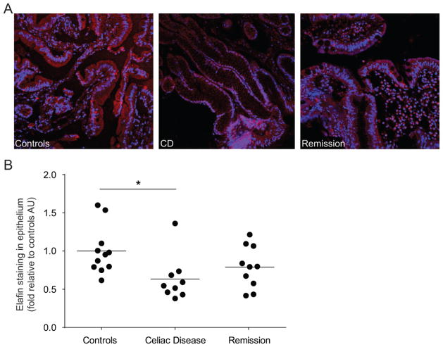
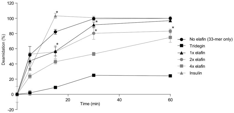
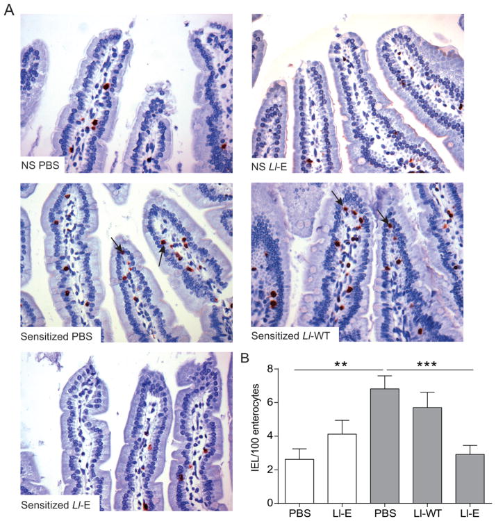
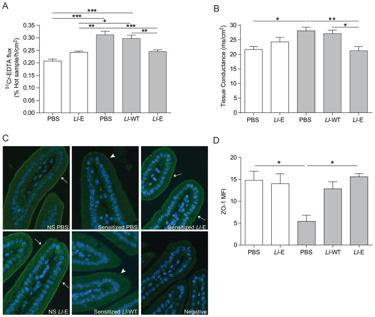
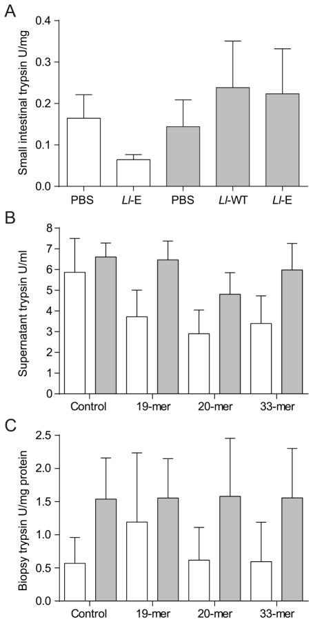
Methods: Epithelial elafin expression in the small intestine of patients with active CD, treated CD, and controls without CD was determined by immunofluorescence. Interaction of elafin with human tissue transglutaminase-2 (TG-2) was investigated in vitro. The 33-mer peptide, a highly immunogenic gliadin peptide, was incubated with TG-2 and elafin at different concentrations. The degree of deamidation of the 33-mer peptide was analyzed by liquid chromatography-mass spectrometry. Elafin was delivered to the intestine of gluten-sensitive mice using a recombinant Lactococcus lactis vector. Small intestinal barrier function, inflammation, proteolytic activity, and zonula occludens-1 (ZO-1) expression were assessed.

Results: Elafin expression in the small intestinal epithelium was lower in patients with active CD compared with control patients. In vitro, elafin significantly slowed the kinetics of the deamidation of the 33-mer peptide to its more immunogenic form. Treatment of gluten-sensitive mice with elafin delivered by the L. lactis vector normalized inflammation, improved permeability, and maintained ZO-1 expression.

Conclusions: The decreased elafin expression in the small intestine of patients with active CD, the reduction of 33-mer peptide deamidation by elafin, coupled to the barrier enhancing and anti-inflammatory effects observed in gluten-sensitive mice, suggest that this molecule may have pathophysiological and therapeutic importance in gluten-related disorders.


Figures

Figure 1
Figure 1
Decreased elafin expression in patients with active celiac disease (CD). Biopsies were obtained from patients in whom CD was excluded (controls), patients with treated CD (1 year gluten-free diet (GFD); remission) and patients with active CD and were stained for elafin (red) expression. (A) Representative immunofluorescence figures are shown. Nuclei labelled with 4′6-diamidino-2-phenylindole (DAPI; blue). (B) Quantification of fluorescence intensity, expressed as a fold increase to control patients. Each dot represents the mean fluorescent value of 4 different areas of one patient and the line represents the mean of each group. *p<0.05.
Figure 2
Figure 2
Deamidation of 33-mer in the competitive reactions. 33-mer peptide (25 μM) was incubated with tissue transglutaminase-2 (TG-2) (1 μM) that served as a control reaction. For the competitive reactions, 25 (x1) or, 50 (x2), or 100 μM (x4) elafin was added. Tridegin and insulin were used as positive and negative controls, respectively. The degree of deamidation was analyzed by liquid chromatography-mass spectrometry (LC-MS). Data are represented as mean ± SEM (n=3). *p<0.05 vs control reaction (no elafin), all data points of the enzymatic reaction with 100 μM (x 4) elafin and tridegin are statistically significant (p<0.02) vs. control reaction (no elafin).
Figure 3
Figure 3
L. lactis expressing elafin(Ll-E) treatment attenuates gliadin-induced intraepithelial lymphocytosis in sensitized NOD/DQ8 mice. (A) CD3+-stained sections of the proximal small intestine. Original magnification x20. Black arrows indicate intraepithelial lymphocytes (IELs). (B) Quantification of CD3+ cells in villi tips, expressed as IEL per 100 enterocytes (n=8–12 per group). White bars represent non-sensitized animals, grey bars represent sensitized animals. Data are represented as mean ± SEM. **p<0.01, ***p<0.001. Non-sensitized (NS); Lactococcus lactis wild type (Ll-WT); L. lactis expressing elafin (Ll-E).
Figure 4
Figure 4
L. lactis expressing elafin(Ll-E) therapy protects NOD/DQ8 mice from gliadin-induced increases in paracellular permeability and preserves zonula occludens-1 (ZO-1) distribution. Sections of small intestine were mounted in Ussing chambers and (A) 51Cr-EDTA flux (% Hot Sample/h/cm2; n=8–11 per group) and (B) tissue conductance (mS/cm2; n=4–7 per group) was measured 24 h after the final gliadin challenge. (C) Sections of the proximal small intestine were stained for ZO-1 (green) expression. Nuclei labelled with 4′6-diamidino-2-phenylindole (DAPI; blue). Original magnification x20. White arrows indicate strong immunofluorescence at the apical junctional complex; arrowheads indicate patchy expression. (D) Mean fluorescence intensity (MFI) of ZO-1 staining in proximal small intestinal sections was determined (n=3–4 per group). MFI was corrected for background fluorescence. White bars represent non-sensitized animals, grey bars represent sensitized animals. Data are represented as mean ± SEM. *p<0.05, **p<0.01, ***p<0.001. Lactococcus lactis wild type (Ll-WT); L. lactis expressing elafin (Ll-E).
Figure 5
Figure 5
Gliadin sensitization and challenge do not alter trypsin-like activity in NOD/DQ8 mice in vivo or in vitro. (A) Sections of proximal small intestine were collected 24 h after final gliadin challenge. Levels of trypsin-like proteases were quantified in tissue homogenates and expressed as U/mg protein (n=8–11 per group). (B, C) Proximal small intestinal biopsies were collected from sensitized and non-sensitized NOD-DQ8 mice and stimulated for 3 h in vitro with gliadin peptides. Trypsin-like activity was quantified in the (B) culture supernatant and (C) biopsies and expressed as U/mL and U/mg protein respectively. White bars represent non-sensitized animals, grey bars represent sensitized animals. Data are represented as mean ± SEM. Lactococcus lactis wild type (Ll-WT); L. lactis expressing elafin (Ll-E).

Comment in

  • Use of elafin in celiac disease.
    Kav T. Kav T. Turk J Gastroenterol. 2015 Jan;26(1):93-4. doi: 10.5152/tjg.2015.0115. Turk J Gastroenterol. 2015. PMID: 25698289 No abstract available.

References

    1. Verdu EF. Editorial: Can Gluten Contribute to Irritable Bowel Syndrome. Am J Gastroenterol. 2011;106:516–8. - PubMed
    1. Verdu EF, Armstrong D, Murray JA. Between celiac disease and irritable bowel syndrome: the “no man’s land” of gluten sensitivity. Am J Gastroenterol. 2009;104:1587–94. - PMC - PubMed
    1. Biesiekierski JR, Newnham ED, Irving PM, et al. Gluten causes gastrointestinal symptoms in subjects without celiac disease: a double-blind randomized placebo-controlled trial. Am J Gastroenterol. 2011;106:508–14. - PubMed
    1. Ludvigsson JF, Leffler DA, Bai JC, et al. The Oslo definitions for coeliac disease and related terms. Gut. 2013;62:43–52. - PMC - PubMed
    1. Pinier M, Fuhrmann G, Galipeau HJ, et al. The copolymer P(HEMA-co-SS) binds gluten and reduces immune response in gluten-sensitized mice and human tissues. Gastroenterology. 2012;142:316–25. - PubMed

Publication types

MeSH terms