. 2012 Sep 25;1(4):667-98.
doi: 10.3390/cells1040667.

The myc road to hearing restoration

Affiliations

The myc road to hearing restoration

Benjamin Kopecky et al. Cells. .

Abstract

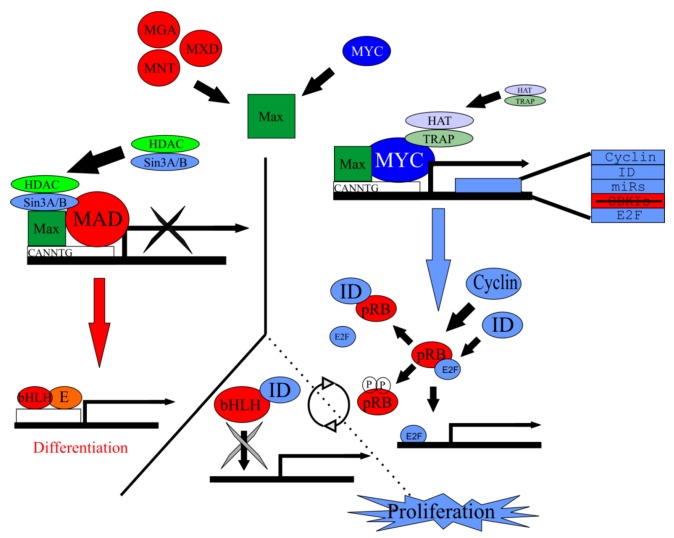
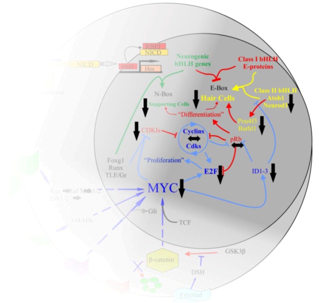
Current treatments for hearing loss, the most common neurosensory disorder, do not restore perfect hearing. Regeneration of lost organ of Corti hair cells through forced cell cycle re-entry of supporting cells or through manipulation of stem cells, both avenues towards a permanent cure, require a more complete understanding of normal inner ear development, specifically the balance of proliferation and differentiation required to form and to maintain hair cells. Direct successful alterations to the cell cycle result in cell death whereas regulation of upstream genes is insufficient to permanently alter cell cycle dynamics. The Myc gene family is uniquely situated to synergize upstream pathways into downstream cell cycle control. There are three Mycs that are embedded within the Myc/Max/Mad network to regulate proliferation. The function of the two ear expressed Mycs, N-Myc and L-Myc were unknown less than two years ago and their therapeutic potentials remain speculative. In this review, we discuss the roles the Mycs play in the body and what led us to choose them to be our candidate gene for inner ear therapies. We will summarize the recently published work describing the early and late effects of N-Myc and L-Myc on hair cell formation and maintenance. Lastly, we detail the translational significance of our findings and what future work must be performed to make the ultimate hearing aid: the regeneration of the organ of Corti.


Figures

Figure 1
Figure 1
Hearing requires the precise stimulation of hair cells. Sound waves from the environment are funneled into the auditory canal by the external ear. Sound waves hit the ear drum and are turned into physical vibrations that are propagated through three middle ear bones, the malleus, the incus, and the stapes. The stapes articulates with the oval window, which separates the middle ear from the inner ear’s fluid-filled scala vestibuli. Physical vibrations of the middle ear bones are converted into compression and rarefaction of the perilymphatic fluid. The scala vestibuli is continuous with the scala tympani. As the fluid waves propagate through the scalae, they cause vibrations of the overlaying basilar membrane. This vibration results in a shearing force of organ of Corti hair cells against the tectorial membrane to activate the auditory nerve (A). Light blue arrows indicate sound wave movement. Black arrows indicate vibration and fluid movement. Dark blue arrows indicate basilar membrane vibration. In conductive hearing loss (B), sound waves are not accurately propagated to the inner ear. Hearing aids (C) can amplify sound waves to overcome this dampening effect. In sensorineural hearing loss (D), hair cells are lost. Hearing aids (E) can partially overcome the resulting hearing deficit by increasing the amplitude to nearby cells; however, no hearing aid can stimulate lost hair cells and furthermore, hearing aids may create discomfort by over-stimulating remaining hair cells. The best current treatment for sensorineural hearing loss is cochlear implants (F) which directly stimulate auditory neurons through an electrode array surgically implanted into the inner ear.
Figure 2
Figure 2
Hearing loss is a major global problem and patient options are limited. Current treatment to restore hearing is centered on the use of auditory prostheses; however, no current option is able to fully restore normal hearing (dashed line). Thus, perfect solutions to this growing problem include prevention of hair cell loss and when hair cell loss does occur, regeneration of damaged tissue is needed to restore full functionality. To successfully prevent the loss of hair cells or regenerate hair cells after loss, we must have a greater understanding of the molecular basis of ear development, specifically, what genes are needed to form and to maintain hair cells. Upon elucidating these minimal essential factors, it may be possible to directly target inner ear tissues. In the case of prevention, increasing the hair cell’s natural maintenance mechanisms may be sufficient. Once hair cells are lost, manipulation of inner ear supporting cells may be possible to form new hair cells. On the other hand, an alternative source of cells, such as stem cells could be first manipulated to form hair cell-like cells and then targeted to the damaged tissue to restore hearing.
Figure 3
Figure 3
Inner ear development requires controlled proliferation. The inner ear begins as an undifferentiated patch of ectoderm adjacent to the hindbrain at embryonic day (E) 8. Through induction by diffusible factors such as the Wnts and Fgfs, neuroectoderm is formed. From this neuroectoderm, neurosensory (NS) precursor cells are formed. NS precursors have the ability to form neurons, hair cells, and supporting cells. An initially small population of NS precursor cells undergoes symmetric division leading to additional NS precursor cells. This multiplication is in part driven by proto-oncogenes such as the Mycs. At a certain point, tumor suppressor genes, pro-sensory genes, and pro-neural genes increase leading to the asymmetric division of NS precursor cells into differentiated NS cells. The formation of NS cells such as neurons and hair cells help maintain the differentiated state of nearby supporting cells. The differentiated state is further maintained by high levels of cyclin dependent kinase inhibitors (CDKIs) and retinoblastoma (Rb). Modified from [33,55,60].
Figure 4
Figure 4
The interactions of Myc in the body are complex. Upon activation of Notch by Jagged or Delta, the Notch IntraCellular Domain (NICD) is cleaved by γ-secretase. NICD translocates to the nucleus to form a complex with RBPJ to positively regulate the transcription of the Hes family of bHLH TFs [63]. Myc is regulated by a number of pathways, including the Wnt/β-catenin pathway, Tgf-β or Bmp/Smad, Fgf/Erk1/2, and the Shh/Smo pathways. Once activated, Myc interacts with partner proteins to regulate a number of downstream effectors, including the cyclins, E2Fs, IDs, CDKIs, and various microRNAs. This regulation leads to the inhibition of class II bHLH TFs from binding to the E-proteins which then allows the Hes family to occupy the E-proteins. It also leads to the removal of pRb from E2F and allows E2F to bind S-phase promoting genes. The larger circle represents the cell as a whole while the smaller circle is the nucleus. Arrows indicate upregulation while blunted lines represent inhibition. Red indicates pathways favoring differentiation, blue indicates pathways favoring proliferation. Modified after [33,55,60,89].
Figure 5
Figure 5
Myc/Max/Mad network regulates proliferation. Max forms a heterodimer with Myc but is also capable of forming a heterodimer with the Mad family of antagonists. Either heterodimer will bind to the CACGTG E-box sequence on target genes. Along with accessory binding partners, Myc/Max will allow for the transcription of target genes responsible for to proliferation. Contrarily, Mad/Max will inhibit the transcription of these target genes, leading ultimately to differentiation. Target genes of Myc/Max include cyclin, ID, E2F, and various microRNAs. Both the cyclins and IDs remove pRb from E2F and allow E2F to bind to S-phase promoting genes and allow subsequent proliferation. IDs are able to bind bHLH TFs but since they lack a basic motif, are unable to bind E-boxes, inhibiting differentiation. In the absence of IDs or cyclins, bHLH TFs are free to bind to E‑proteins and the E-box, promoting differentiation. Modified from [89].
Figure 6
Figure 6
The Myc interactions in the inner ear require additional clarification. We began our exploration of N-Myc and L-Myc function following our stated hypothesis that N-Myc and L-Myc were important to proliferation and possibly followed similar pathways in the inner ear as the Mycs had been described to elsewhere in the body. While the full mechanisms require further elucidation, we have provided some clarity to the picture. N‑Myc, and to a lesser extent, L-Myc are important to inner ear development. In the absence of Myc, E2F, ID, CDKI, Atoh1, ND1, Pou4f3, and Barhl1 are all reduced. cyclinD2 and pRb appear not to be greatly affected. This leads to a progressive loss of the organ of Corti hair cells. Figure modified from [55,60,89].

Similar articles

Cited by

References

    1. Stevens G., Flaxman S., Brunskill E., Mascarenhas M., Mathers C.D., Finucane M. Global and regional hearing impairment prevalence: An analysis of 42 studies in 29 countries. Eur. J. Public Health. 2011 - PubMed
    1. Pauley S., Kopecky B., Beisel K., Soukup G., Fritzsch B. Stem cells and molecular strategies to restore hearing. Panminerva Med. 2008;50:41–53. - PMC - PubMed
    1. Corwin J.T., Cotanche D.A. Regeneration of sensory hair cells after acoustic trauma. Science. 1988;240:1772–1774. - PubMed
    1. Ryals B.M., Rubel E.W. Hair cell regeneration after acoustic trauma in adult Coturnix quail. Science. 1988;240:1774–1776. - PubMed
    1. OTOscope. [(accessed on 19 June 2012)]. Available online: http://www.healthcare.uiowa.edu/labs/morl/otoscope/home.html.

LinkOut - more resources