

doi: 10.1038/cr.2014.44.
Epub 2014 Apr 8.
Double-stranded DNA in exosomes: a novel biomarker in cancer detection
Affiliations
- PMID: 24710597
- PMCID: PMC4042169
- DOI: 10.1038/cr.2014.44
Item in Clipboard
Double-stranded DNA in exosomes: a novel biomarker in cancer detection
Cell Res.
2014 Jun.
No abstract available
Figures
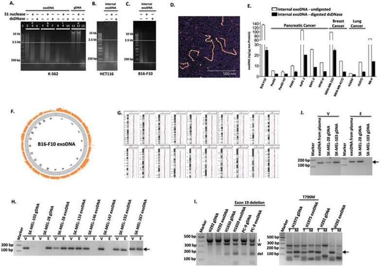
Identification and characterization of exoDNA and its potential use as a diagnostic tool. (A) Equal amounts of DNA extracted from K-562 exosomes, which were untreated (Set a), pre-treated with S1 nuclease (Set b) or dsDNase (Set c), were digested with either S1 nuclease (lanes 3, 6 and 9) or dsDNase (lanes 4, 7 and 10). Digestion of gDNA (Set d) with S1 nuclease (lane 12) or dsDNase (lane 13) serves as controls. (B, C) Analysis of internal exoDNA isolated from HCT116 (B) and B16-F10 (C) after removal of external exoDNA as in A. The results are representative of 2-3 experiments performed independently. (D) AFM image of exoDNA. ExoDNA was extracted from K-562 cells and absorbed on a mica surface in the presence of 5 mM Mg2+. Scale bar, 500 nm. (E) Internal exoDNA was extracted from exosomes secreted by different types of cancer cell lines, including melanoma (B16-F10), pancreatic cancer (Pan02, Pan02 H3, PANC-1, AsPC-1, BXPC-3 and HPAF-II), breast cancer (MDA-MB-231 and MDA-MB-4175), lung cancer (H1650 and H1975), and leukemia (NB-4), and digested with dsDNase. Abundance of dsDNA inside the exosomes, before and after digestion with dsDNase, was expressed as “nanogram of DNA per microgram of exo-Protein”. (F) Circular view of the readings of fragments along each chromosome in the whole-genome sequencing analysis of exoDNA isolated from murine melanoma B16-F10 cell-derived exosomes. (G) ExoDNA represents gDNA shown by comparative genomic hybridization array analysis of B16-F10 exoDNA vs gDNA. (H-J) Mutational analysis of exoDNA. BRAF(V600E) mutation in exoDNA isolated from either cultured melanoma cell lines (H) or circulating exoDNA isolated from SK-MEL-28 melanoma-bearing mice (J) was detected by AS-PCR analysis. gDNA isolated from SK-MEL-28 and SK-MEL-103 cells serves as a positive and negative control, respectively, for V600E mutation (WT (V) and mutant (E) alleles). Arrow indicates the size of expected PCR products. AS-PCR analysis of EGFR mutations in exoDNA isolated from NSCLC cells was shown in I. For del19, “I” indicates internal control; “W”, WT; and “del”, deletion of exon 19. For T790M mutation, “T” indicates WT allele and “M” indicates the mutant allele. The arrow marks the expected size of PCR products.
References
Publication types
MeSH terms
Substances
Grants and funding
LinkOut - more resources
Full Text Sources
Other Literature Sources
