. 2015 Aug;37(8):1088-95.
doi: 10.1002/hed.23706. Epub 2014 Sep 16.

Expression profile and in vitro blockade of programmed death-1 in human papillomavirus-negative head and neck squamous cell carcinoma

Affiliations

Expression profile and in vitro blockade of programmed death-1 in human papillomavirus-negative head and neck squamous cell carcinoma

Ian-James Malm et al. Head Neck. 2015 Aug.

Abstract

Background: Treatment with a blocking programmed death-1 (αPD-1) antibody recently showed clinical efficacy for various tumor types. In this study, we characterized the expression profile of PD-1/programmed death-ligand-1 (PD-L1) and the potential of PD-1 blockade in human papillomavirus (HPV)-negative head and neck squamous cell carcinoma (HNSCC).

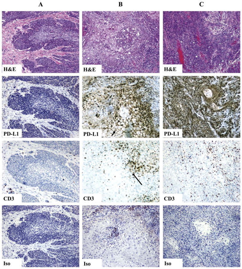
Methods: Lymphocytes from peripheral blood, draining lymph nodes, and the tumor were phenotyped for PD-1 expression, and their proliferative activity was assessed in the presence of blocking αPD-1 treatment. Primary tumor expression of PD-L1 was also analyzed using immunohistochemistry (IHC).

Results: Lymphocyte PD-1 expression was abundant with highest expression in the tumor, and in vitro mixed lymphocyte reaction demonstrated that PD-1 blockade could induce T cell proliferation. Furthermore, tumor cells were found to have 3 distinct patterns of PD-L1 expression with over 78% of the specimens demonstrating strong PD-L1 positivity.

Conclusion: Our data strongly supports the use of αPD-1 blockade in patients with HPV-negative HNSCC that are refractory to standard treatments.

Keywords: T cell; anergy; head and neck squamous cell carcinoma (HNSCC); immunotherapy; programmed death-1 (PD-1).


Figures

FIGURE 1
FIGURE 1
Programmed death-1 (PD-1) and lymphocyte-activation gene 3 (LAG-3) expression on T cells from patients with head and neck squamous cell carcinoma (HNSCC). (A) CD4 and CD8 T cells isolated from peripheral blood, draining lymph node, or tumor were isolated and stained for PD-1 and LAG-3 expression. Cells were gated on CD4 and CD8 T cells before analysis of checkpoint molecule expression. (B) Synopsis of PD-1 and LAG-3 expression on T cells in patients with HNSCC (n = 4 – 11, respectively).
FIGURE 2
FIGURE 2
In vitro programmed death-1 (PD-1) blockade enhances draining lymph node CD4 and CD8 T cell function in patients with head and neck squamous cell carcinoma (HNSCC). (A) Synopsis of proliferation in CD4 and CD8 T cells in a mixed lymphocyte reaction (n = 4). (B) Synopsis of interferon-gamma (IFN-γ) production from CD4 and CD8 T cells in a mixed lymphocyte reaction (n = 4).
FIGURE 3
FIGURE 3
Draining lymph node CD4 and CD8 T-cell function is augmented by the addition of interleukin (IL)-2 alone in patients with head and neck squamous cell carcinoma (HNSCC). (A) Synopsis of proliferation in CD4 and CD8 T cells in a mixed lymphocyte reaction (n = 4). (B) Synopsis of interferon-gamma (IFN-γ) production from CD4 and CD8 T cells in a mixed lymphocyte reaction (n = 4).
FIGURE 4
FIGURE 4
Programmed death-1 (PD-1)high CD8 cells can be stimulated to expresses interferon-gamma (IFN-γ). Synopsis of PMA/ionomycin stimulation of CD8 T cells (n = 2). CD3+ cells were sorted based on PD-1 expression, stimulated, and then stained for CD8 and IFN-γ.
FIGURE 5
FIGURE 5
Human papillomavirus (HPV)-negative head and neck squamous cell carcinoma (HNSCC) tumors express programmed death ligand-1 (PD-L1). Tumor samples from the oropharynx and larynx were sectioned and stained for PD-L1 and CD3. Three patterns of staining were observed: (A) absence of PD-L1 staining with few lymphocytes, (B) regional expression of PD-L1 (membranous “chicken-wire” staining, arrow) colocalized with invading CD3+ lymphocytes (diamond-arrow), and (C) out of proportion PD-L1 staining with rare/sporadic lymphocytes (original magnification ×10). [Color figure can be viewed in the online issue, which is available at wileyonlinelibrary.com.]

References

    1. Pulte D, Brenner H. Changes in survival in head and neck cancers in the late 20th and early 21st century: a period analysis. Oncologist. 2010;15:994–1001. - PMC - PubMed
    1. Davidson HC, Leibowitz MS, Lopez–Albaitero A, Ferris RL. Immunotherapy for head and neck cancer. Oral Oncol. 2009;45:747–751. - PMC - PubMed
    1. Dougan M, Dranoff G. Immune therapy for cancer. Annu Rev Immunol. 2009;27:83–117. - PubMed
    1. Pardoll DM. The blockade of immune checkpoints in cancer immunotherapy. Nat Rev Cancer. 2012;12:252–264. - PMC - PubMed
    1. Drake CG, Jaffee E, Pardoll DM. Mechanisms of immune evasion by tumors. Adv Immunol. 2006;90:51–81. - PubMed

Publication types

MeSH terms