

Review
doi: 10.1109/TBME.2014.2314733.
Epub 2014 Apr 2.
Retinal prosthesis
- PMID: 24710817
- PMCID: PMC4356127
- DOI: 10.1109/TBME.2014.2314733
Item in Clipboard
Review
Retinal prosthesis
IEEE Trans Biomed Eng.
2014 May.
Abstract
Retinal prosthesis has been translated from the laboratory to the clinic over the past two decades. Currently, two devices have regulatory approval for the treatment of retinitis pigmentosa. These devices provide partial sight restoration and patients use this improved vision in their everyday lives. Improved mobility and object detection are some of the more notable findings from the clinical trials. However, significant vision restoration will require both better technology and improved understanding of the interaction between electrical stimulation and the retina. This paper reviews the recent clinical trials and highlights technology breakthroughs that will contribute to next generation of retinal prostheses.
Figures

Retinal Prosthesis Concept. An image is converted to an electrical signal by an imaging device. The electrical signal is processed by external and/or implanted circuitry. The circuit produces a pattern of electrical stimulus which is applied to the retina via a microelectrode array positioned near the retina. Image courtesy of Annual Review of Biomedical Engineering.

The Argus II Retina Prosthesis. A. The external system has a camera mounted in a pair of glasses. The video processing unit (VPU) processes the camera data, then transmits wireless power and data via the coil. B. The implant coil receives power and data for processing by the electronics (within the silver case). The coil and case are secured with a scleral band that encircles the eye. The electrode cable traverses the eye wall and the electrode array is attached to the array with a retinal tack. Images from [12]
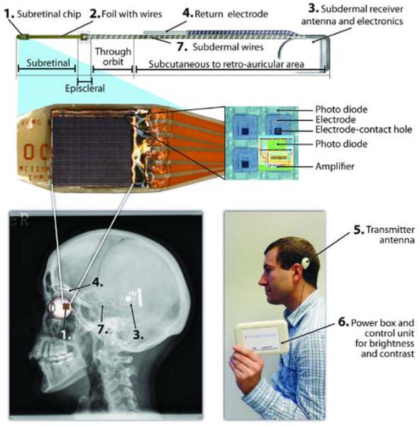
Alpha – IMS system (Retina Implant, GmbH). 1. The subretinal photodiode array (MPDA). The MPDA has 1500 elements, each of which has a photodiode to sense light, circuitry to amplify the signal, and electrode to deliver the stimulus pulse. 2. A micro flex cable holds the MPDA while larger wire connect to 3. the subdermal receiver. 4. The return electrode is outside the eye, under the skin. 5 & 6. The external system provides power to the implant as well as some configuration data. 7. The implant position on the head. Images Courtesy Dr. Eberhart Zrenner.

Hermetic Packaging Schemes. Top – An electronics modules is placed inside an enclosure, which includes a feedthrough platform with conductors and a case or lid. The feedthrough and case are sealed together. Bottom – Coating an electronic chip is an ideal protection scheme, but to date no coating technology has proven adequate for long-term implantation.
High density feedthrough technology. Front view shows platinum conductors embedded in alumina substrate.
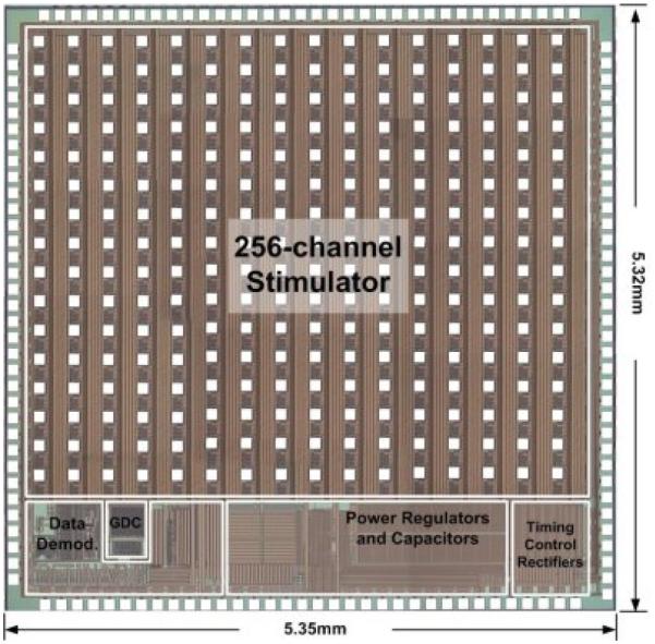
A 256 channel stimulator by Liu and colleagues. The chip has integrated power conditioning and data decode, and 256 independent output channels. Image courtesy of Dr. Wentai Liu.
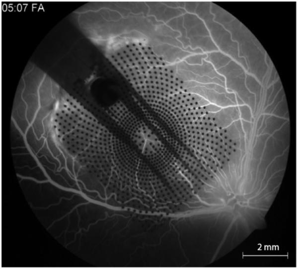
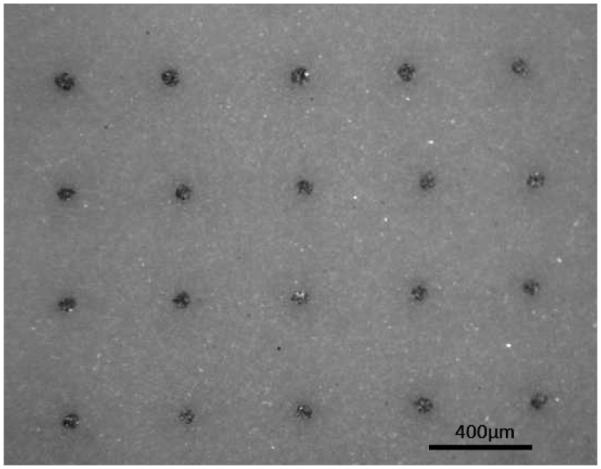
A parylene substrate electrode array with 1000 individual electrode contacts, implanted in the eye of a canine. The image is acquired via a fundus camera, through the dilated pupil. Fluorscein injected into the circulatory systemically shows integrity of the retinal blood vessels under the array.

An intraocular camera positioned in place of the crystalline lens, similar to what is done in cataract surgery when an intraocular lens is implanted. Such a device will allow eye gaze to be in accord with perception, a key to improving the visual capability of implant patients. Image courtesy Dr. Armand R. Tanguay, Jr.

PET imaging of (left) Light stimulation in subjects with normal vision and (right) Corneal electrical stimulation in blind subjects (with RP). Posterior view shown, arrows indicate posterior pole. Courtesy Dr. John Xie.
References
-
- Dowling JE. The Retina: an approachable part of the Brain. Bellknap Press; 1987.
-
- Hartong DT, Berson EL, Dryja TP. Retinitis Pigmentosa. Lancet. 2006 Nov;368:1795–1809. - PubMed
-
- Potts AM, Inoue J. The electrically evoked response (EER) of the visual system. II. Effect of adaptation and retinitis pigmentosa. Invest. Ophth. 1969 Jun;8(6):605–12. - PubMed
Publication types
MeSH terms
Grants and funding
LinkOut - more resources
Full Text Sources
Other Literature Sources
Miscellaneous
