

Mutual information as a measure of image quality for 3D dynamic lung imaging with EIT
- PMID: 24710978
- PMCID: PMC4059506
- DOI: 10.1088/0967-3334/35/5/863
Mutual information as a measure of image quality for 3D dynamic lung imaging with EIT
Abstract
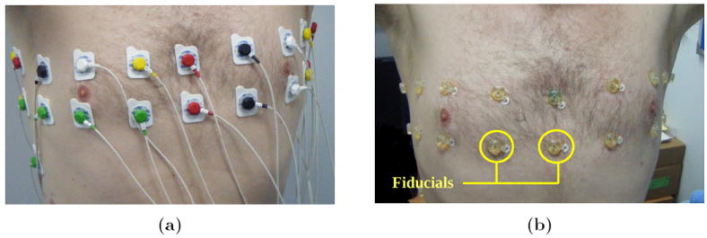
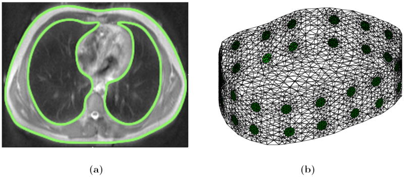
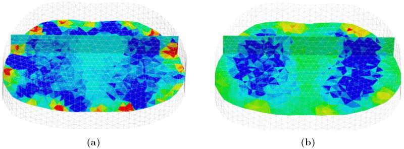
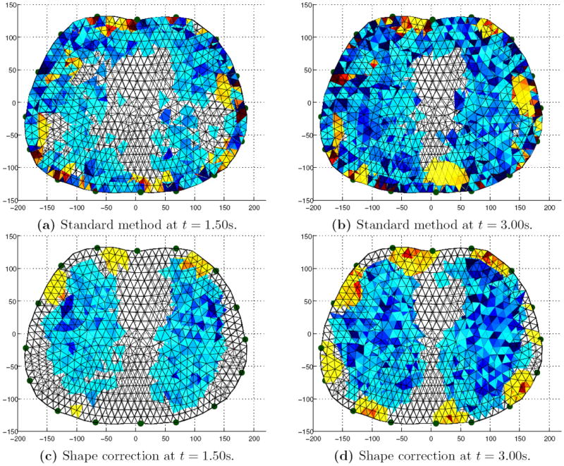
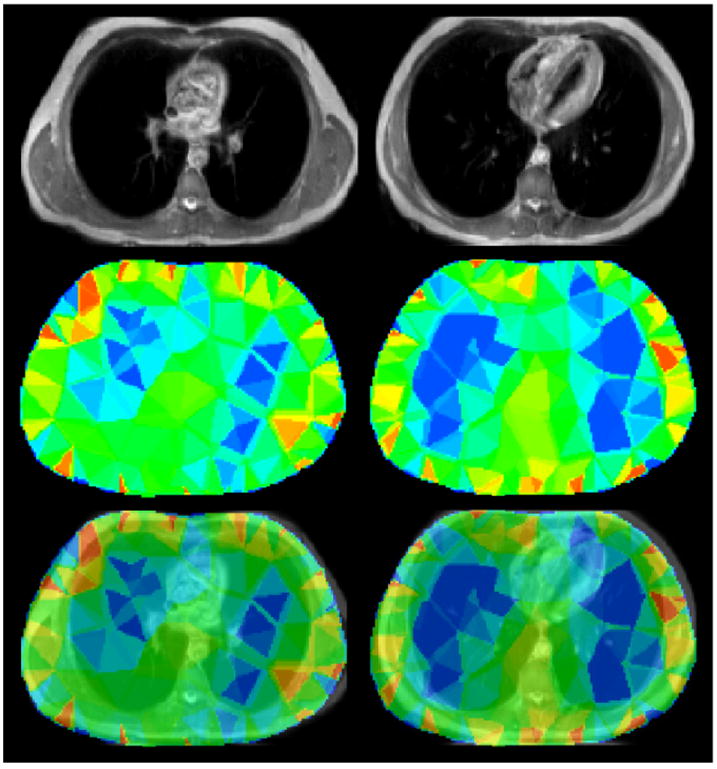
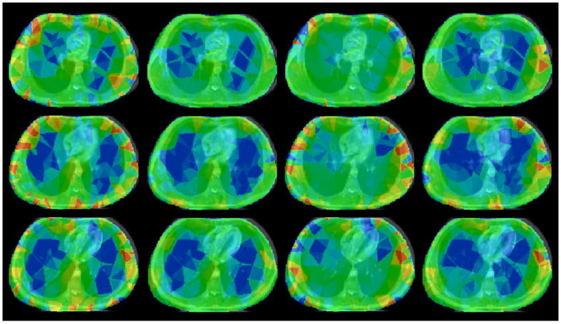
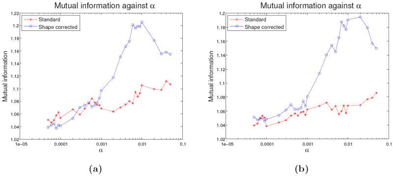
We report on a pilot study of dynamic lung electrical impedance tomography (EIT) at the University of Manchester. Low-noise EIT data at 100 frames per second were obtained from healthy male subjects during controlled breathing, followed by magnetic resonance imaging (MRI) subsequently used for spatial validation of the EIT reconstruction. The torso surface in the MR image and electrode positions obtained using MRI fiducial markers informed the construction of a 3D finite element model extruded along the caudal-distal axis of the subject. Small changes in the boundary that occur during respiration were accounted for by incorporating the sensitivity with respect to boundary shape into a robust temporal difference reconstruction algorithm. EIT and MRI images were co-registered using the open source medical imaging software, 3D Slicer. A quantitative comparison of quality of different EIT reconstructions was achieved through calculation of the mutual information with a lung-segmented MR image. EIT reconstructions using a linear shape correction algorithm reduced boundary image artefacts, yielding better contrast of the lungs, and had 10% greater mutual information compared with a standard linear EIT reconstruction.
Figures
References
-
- Adler A, et al. GREIT: a unified approach to 2D linear EIT reconstruction of lung images. Physiol Meas. 2009;30:S35–55. - PubMed
-
- Adler A, et al. Whither lung EIT: where are we, where do we want to go and what do we need to get there? Physiol Meas. 2012;33:679–94. - PubMed
-
- Adler A, Borsic A, Polydorides A, Lionheart WRB. Simple FEMs aren’t as good as we thought: experiences developing EIDORS v3.3. Proc 9th Int Conf EIT; Dartmouth College, New Hampshire. 2008.
-
- Adler A, Guardo R, Berthiaume Y. Impedance imaging of lung ventilation: do we need to account for chest expansion? IEEE Trans Biomed Eng. 1996;43:414–20. - PubMed
-
- Adler A, Lionheart WRB. Uses and abuses of EIDORS: An extensible software base for EIT. Physiol Meas. 2006;27:S25–42. - PubMed
Publication types
MeSH terms
Grants and funding
LinkOut - more resources
Full Text Sources
Other Literature Sources