. 2014 Apr 22;111(16):6075-80.
doi: 10.1073/pnas.1402251111. Epub 2014 Apr 7.

Cholinergic neurotransmission links solitary chemosensory cells to nasal inflammation

Affiliations

Cholinergic neurotransmission links solitary chemosensory cells to nasal inflammation

Cecil J Saunders et al. Proc Natl Acad Sci U S A. .

Abstract

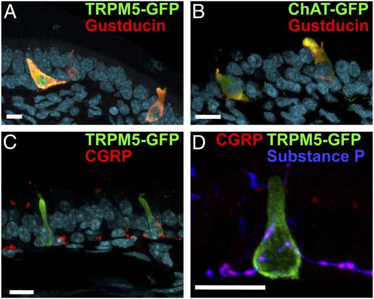
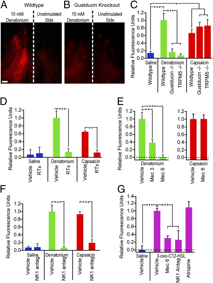
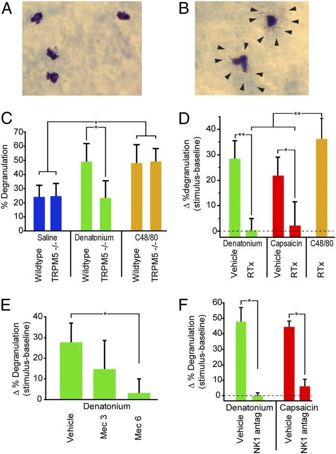
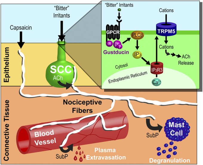
Solitary chemosensory cells (SCCs) of the nasal cavity are specialized epithelial chemosensors that respond to irritants through the canonical taste transduction cascade involving Gα-gustducin and transient receptor potential melastatin 5. When stimulated, SCCs trigger peptidergic nociceptive (or pain) nerve fibers, causing an alteration of the respiratory rate indicative of trigeminal activation. Direct chemical excitation of trigeminal pain fibers by capsaicin evokes neurogenic inflammation in the surrounding epithelium. In the current study, we test whether activation of nasal SCCs can trigger similar local inflammatory responses, specifically mast cell degranulation and plasma leakage. The prototypical bitter compound, denatonium, a well-established activator of SCCs, caused significant inflammatory responses in WT mice but not mice with a genetic deletion of elements of the canonical taste transduction cascade, showing that activation of taste signaling components is sufficient to trigger local inflammation. Chemical ablation of peptidergic trigeminal fibers prevented the SCC-induced nasal inflammation, indicating that SCCs evoke inflammation only by neural activity and not by release of local inflammatory mediators. Additionally, blocking nicotinic, but not muscarinic, acetylcholine receptors prevents SCC-mediated neurogenic inflammation for both denatonium and the bacterial signaling molecule 3-oxo-C12-homoserine lactone, showing the necessity for cholinergic transmission. Finally, we show that the neurokinin 1 receptor for substance P is required for SCC-mediated inflammation, suggesting that release of substance P from nerve fibers triggers the inflammatory events. Taken together, these results show that SCCs use cholinergic neurotransmission to trigger peptidergic trigeminal nociceptors, which link SCCs to the neurogenic inflammatory pathway.

Keywords: airway irritation; chemesthesis; innate immunity; quorum sensing; rhinitis.


Conflict of interest statement

The authors declare no conflict of interest.

Figures

Fig. 1.
Fig. 1.
Cross-sections of nasal epithelium showing cellular properties and relationships of SCCs. (A) SCCs express both TRPM5 (detected by TRPM5-driven GFP; green) and gustducin (red), which are elements of the canonical taste transduction pathway. (B) SCCs express both gustducin (red) and ChAT (detected by ChAT-driven GFP; green), the synthesizing enzyme for ACh. (C) SCCs expressing TRPM5 (green) are intimately contacted by calcitonin gene-related peptide (CGRP) immunoreactive peptidergic nociceptive trigeminal fibers. (D) These peptidergic fibers (magenta) contact SCCs (TRPM5-GFP; green) and are immunoreactive for both CGRP (red) and substance P (blue). The nuclear counterstain DRAQ5 is shown in cyan in A–C. (Scale bars: 10 µm.)
Fig. 2.
Fig. 2.
Stimulation of SCCs or nociceptor nerve terminals activates a proinflammatory pathway, leading to plasma extravasation. (A and B) Fluorescence images of a whole mount of the hemisected nasal cavity from mice stimulated unilaterally with 10 mM denatonium benzoate and injected i.v. with Alexa 555-albumin showing fluorescence because of plasma leakage. Anterior is up. (Scale bar: 1 mm.) (A) WT mouse shows increased fluorescence on the stimulated side. (B) A Gustducin−/− mouse shows no significant fluorescence on either the stimulated or unstimulated sides. (C–G) Bar graphs illustrating the relative fluorescence of stimulated and unstimulated sides under various conditions and genotypes. Positive values indicate that the stimulated side was brighter than the unstimulated side. Bars represent mean + SEM. (C) WT mice stimulated with 10 mM denatonium (green) or 2 μM capsaicin (red) showed significant (P < 0.001 or P < 0.01, respectively) plasma extravasation on the stimulated side compared with saline-stimulated mice (blue). Two KO strains, Gustducin−/− and TRPM5−/−, showed significantly less (P < 0.001) extravasation than WT controls stimulated with denatonium but normal extravasation with capsaicin. (D) Mice treated with RTx to eliminate peptidergic nerve fibers were significantly different from vehicle-treated controls (P < 0.01 and P < 0.001) and showed no significant extravasation to either denatonium or capsaicin. (E) The nAChR antagonist mecamylamine (Mec) significantly reduced denatonium-induced extravasation at both 3 (P < 0.01) and 6 mg/kg (P < 0.001) but did not alter capsaicin-induced extravasation. (F) The NK1 antagonist L732138 (5 mg/kg i.p.), which blocks responses to substance P, significantly reduces plasma extravasation in response to both denatonium (P < 0.001) and capsaicin (P < 0.001). (G) Stimulation with the bacterial metabolite 3-oxo-C12-HSL (300 μM) provoked significant (P < 0.001) plasma extravastion compared with saline-stimulated controls. This HSL-induced plasma extravasation was significantly reduced (P < 0.001) by treatment with either the nicotinic antagonist mecamylamine or the NK1 antagonist L732138, but was not altered by the muscarinic AChR blocker atropine (1 mg/kg). *P < 0.05; **P < 0.01; ***P < 0.001 by one-way ANOVA with Tukey honest significant difference (HSD) test.
Fig. 3.
Fig. 3.
Stimulation of SCCs activates a proinflammatory pathway that triggers mast cell degranulation. Photos of (A) resting and (B) degranulated mast cells stained with acidified toluidine blue. Arrows point to granules forming a halo around the degranulated mast cells. (C) WT mice stimulated with 10 mM denatonium showed significantly more mast cell degranulation than TRPM5−/− animals (P < 0.05). Both WT and TRPM5−/− mice showed degranulation on exposure to the secretagague C48/80. (D) Mice treated with RTx to eliminate nerve fibers showed significantly less mast cell degranulation than vehicle-treated controls to both denatonium (P < 0.001) and capsaicin (P < 0.01) but were still able to respond to compound 48/80 (C48/80), which directly acts on mast cells to cause degranulation. (E) The nAChR antagonist mecamylamine (Mec) significantly reduced denatonium-induced mast cell degranulation at a dose of 6 mg/kg (P < 0.01). (F) The NK1 antagonist L732138 (5 mg/kg i.p.), which blocks responses to substance P, significantly reduces mast cell degranulation in response to both denatonium (P < 0.05) and capsaicin (P < 0.05). Bars represent mean + SEM. *P < 0.05; **P < 0.01 by one-way ANOVA with Tukey HSD test.
Fig. 4.
Fig. 4.
Parallel pathways for airway irritation. SCCs (green) respond to bitter substances, such as denatonium and bacterial metabolites, through the canonical taste transduction pathway. SCCs release ACh, which activates nAChRs on nociceptive trigeminal nerve fibers (white) that innervate the SCCs. These nociceptive trigeminal fibers also express TRPV1 and TRPA1, two chemosensitive ion channels that are responsive to irritants, such as capsaicin. Regardless of whether the nociceptive fiber is activated directly through TRP channels or indirectly through SCCs, the collateral terminals release inflammatory mediators. One of these mediators, substance P (SubP), activates NK1 receptors on blood vessels (red), resulting in plasma extravasation, and mast cells (blue), causing degranulation. (Inset) Irritants stimulate G protein-coupled receptors (GPCRs) to activate the βγ-subunit associated with α-gustducin, thereby producing inositiol trisphosphate (IP3) through a phospholipase Cβ2 (PLCβ2) -mediated cascade. In turn, IP3 binds to the type 3 IP3 receptor (IP3R3), releasing Ca2+ from the endoplasmic reticulum. Increases in cytosolic Ca2+ activate TRPM5, a nonspecific cation channel, and lead to depolarization and release of ACh.

References

    1. Silver W, Roe P, Saunders CJ. Functional neuroanatomy of the upper airway in experimental animals. In: Morris JB, Shusterman D, editors. Toxicology of the Nose and Upper Airways. New York: Informa Healthcare; 2010. pp. 45–64.
    1. Silver WL, Finger TE. The anatomical and electrophysiological basis of peripheral nasal trigeminal chemoreception. Ann N Y Acad Sci. 2009;1170:202–205. - PubMed
    1. Bryant BP, Silver W. Chemesthesis: The common chemical sense. In: Finger TE, Silver W, Restrepo D, editors. Neurobiology of Taste and Smell. 2nd Ed. New York: Wiley-Liss; 2000. pp. 73–100.
    1. Kobayashi K, et al. Distinct expression of TRPM8, TRPA1, and TRPV1 mRNAs in rat primary afferent neurons with adelta/c-fibers and colocalization with trk receptors. J Comp Neurol. 2005;493(4):596–606. - PubMed
    1. Finger TE, St Jeor VL, Kinnamon JC, Silver WL. Ultrastructure of substance P- and CGRP-immunoreactive nerve fibers in the nasal epithelium of rodents. J Comp Neurol. 1990;294(2):293–305. - PubMed

Publication types

MeSH terms