. 2014 May 23;289(21):14534-46.
doi: 10.1074/jbc.M114.564674. Epub 2014 Apr 7.

Smoke exposure causes endoplasmic reticulum stress and lipid accumulation in retinal pigment epithelium through oxidative stress and complement activation

Affiliations

Smoke exposure causes endoplasmic reticulum stress and lipid accumulation in retinal pigment epithelium through oxidative stress and complement activation

Kannan Kunchithapautham et al. J Biol Chem. .

Abstract

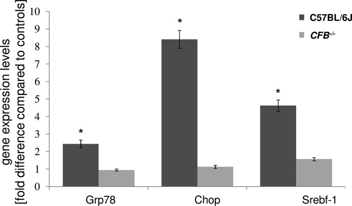
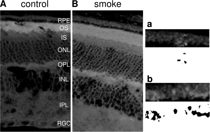
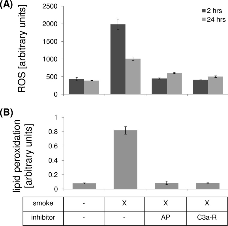
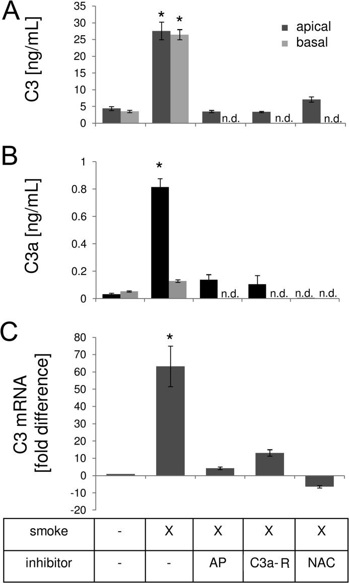
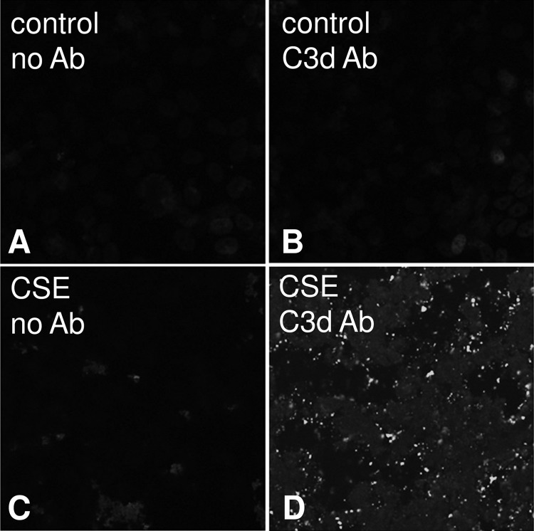
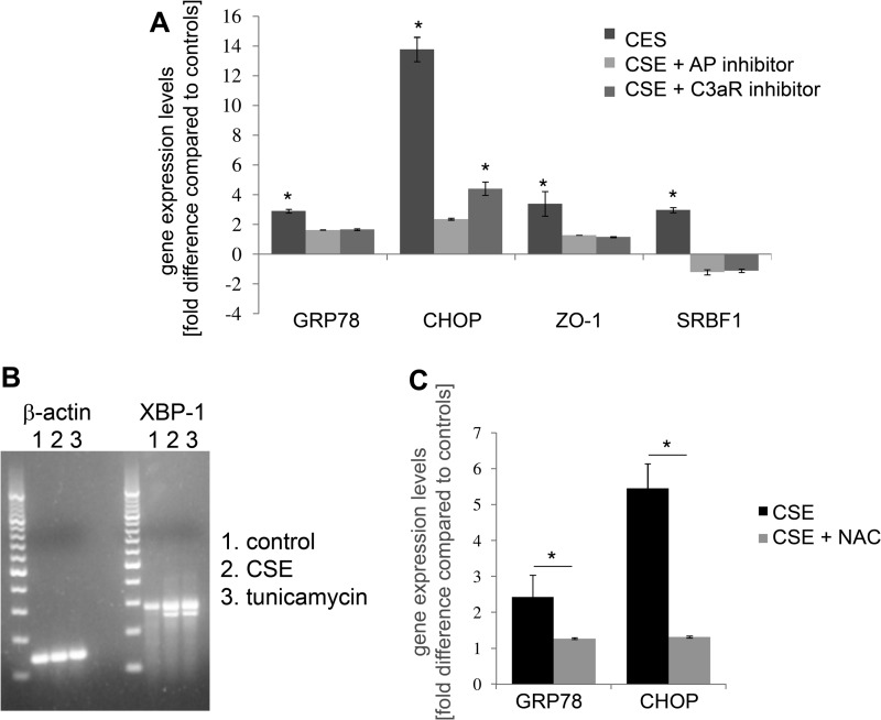
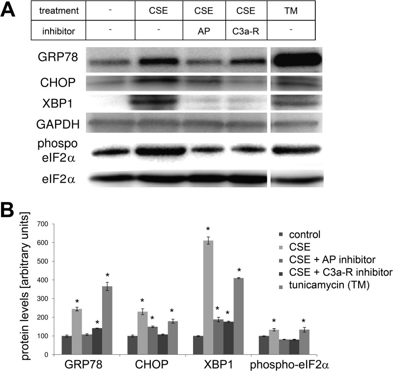
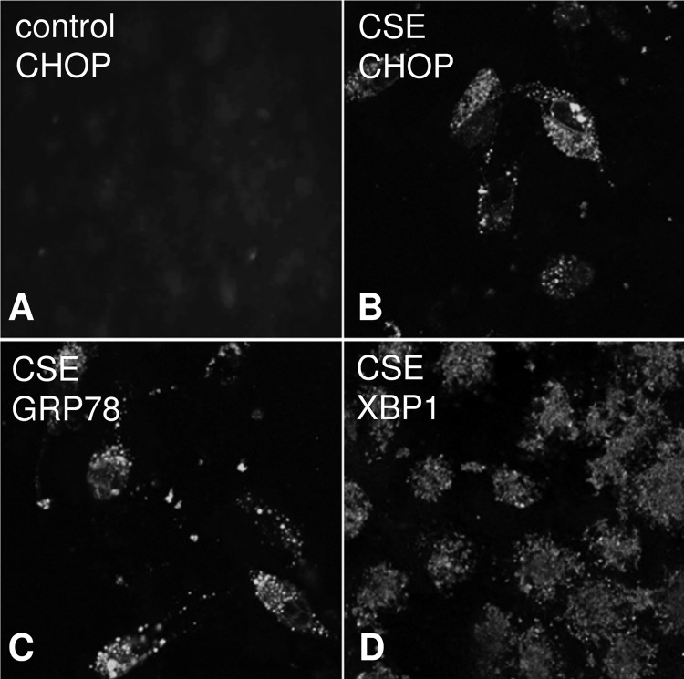
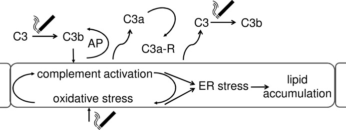
Age-related macular degeneration (AMD) is a complex disease caused by genetic and environmental factors, including genetic variants in complement components and smoking. Smoke exposure leads to oxidative stress, complement activation, endoplasmic reticulum (ER) stress, and lipid dysregulation, which have all been proposed to be associated with AMD pathogenesis. Here we examine the effects of smoke exposure on the retinal pigment epithelium (RPE). Mice were exposed to cigarette smoke or filtered air for 6 months. RPE cells grown as stable monolayers were exposed to 5% cigarette smoke extract (CSE). Effects of smoke were determined by biochemical, molecular, and histological measures. Effects of the alternative pathway (AP) of complement and complement C3a anaphylatoxin receptor signaling were analyzed using knock-out mice or specific inhibitors. ER stress markers were elevated after smoke exposure in RPE of intact mice, which was eliminated in AP-deficient mice. To examine this relationship further, RPE monolayers were exposed to CSE. Short term smoke exposure resulted in production and release of complement C3, the generation of C3a, oxidative stress, complement activation on the cell membrane, and ER stress. Long term exposure to CSE resulted in lipid accumulation, and secretion. All measures were reversed by blocking C3a complement receptor (C3aR), alternative complement pathway signaling, and antioxidant therapy. Taken together, our results provide clear evidence that smoke exposure results in oxidative stress and complement activation via the AP, resulting in ER stress-mediated lipid accumulation, and further suggesting that oxidative stress and complement act synergistically in the pathogenesis of AMD.

Keywords: Age-related Macular Degeneration; Complement System; ER Stress; Lipodystrophy; Mitochondria; Oxidative Stress; Smoking; Unfolded Protein Response.


Figures

FIGURE 1.
FIGURE 1.
Gene expression changes in ocular tissues between WT and CFB−/− mice following CSE. Analysis of marker gene expression in WT and CFB−/− mice, using quantitative RT-PCR on cDNA generated from a RPE/choroid/sclera fraction. Quantitative values were obtained by cycle number (Ct value), determining the difference between the mean experimental and control (β-actin) ΔCt values for smoke versus room air-exposed mice within each genotype (fold-difference). Candidates were examined from two categories, ER stress (Grp78 and Chop) and lipid metabolism (Srebf-1). Significant changes were identified in all three genes for C57BL/6J mice, whereas gene expression was minimally affected in CFB−/− animals. Data are expressed as mean ± S.E. (n = 3 per condition, *, p < 0.05).
FIGURE 2.
FIGURE 2.
Lipid deposition in eyes exposed to smoke. Localization of neutral lipids was identified using LipidTox staining, comparing C57BL/6J mice exposed to room air (A) or smoke (B). Lipid particles were seen in Bruch's membrane (BrM) and the basal side of the RPE in smoke-exposed mice when compared with controls. Insets highlight RPE/BrM and corresponding binarized images from control and smoke-exposed eyes (a and b). Abbreviations: RPE, retinal pigment epithelium; OS, outer segments; IS, inner segments; ONL, outer nuclear layer; OPL, outer plexiform layer; INL, inner nuclear layer; IPL, inner plexiform layer; RGC, retinal ganglion cell layer.
FIGURE 3.
FIGURE 3.
Lipid deposition in ARPE-19 cell monolayers exposed to smoke. Localization of lipids was identified using Nile Red (A, B, and F–I), and unesterified (C and D) and esterified cholesterol (E) was characterized, comparing cells exposed to control media (A and C) or smoke (B and D–G). Nile Red-positive lipid particles were seen in RPE cells after smoke exposure (B) when compared with controls (A). Lipid deposits could also be detected on the filters of the Transwell plates after cells were completely detached, confirming basolateral release of lipids from these cells (b, inset). Lipid particles contained both unesterified (D) and esterified cholesterol (E). Nile Red-positive lipid particles were eliminated in cells pretreated with the alternative pathway inhibitor CR2-fH (F) or specific C3a-receptor antagonist (G) for 1 h prior to smoke exposure. Monolayers treated daily with tunicamycin to induce ER stress were found to stain strongly for Nile Red-positive lipids (H); levels that did not change after pretreatment with CR2-fH (I).
FIGURE 4.
FIGURE 4.
Oxidative stress in ARPE-19 cells exposed to smoke. Cells were grown into mature monolayers, exposed to CSE, and collected for analysis after 2 or 24 h. Some wells were pretreated with CR2-fH or C3a-receptor antagonist for 1 h. A, ROS were determined using DCFDA. Levels were elevated by CSE after 2 or 24 h and reduced after complement inhibition. B, oxidative degradation of lipids was determined by examining lipid peroxidation by measuring the formation of MDA, a natural side product of this process. After 2 h, levels were elevated by CSE and reduced after complement inhibition. Data are expressed as mean ± S.E. (n = 3 per condition, *, p < 0.05).
FIGURE 5.
FIGURE 5.
Complement activation in ARPE-19 cells exposed to smoke. Cells were grown into mature monolayers, exchanged to serum-free media 24 h prior to the experiment, exposed to CSE in serum-free media, and collected for ELISA analysis after 4 h, or quantitative RT-PCR after 24 h. A and B, ELISA was performed, using plates coated with C3- or C3a-specific capture antibodies. Some wells were pretreated with CR2-fH, C3a-receptor antagonist, or antioxidant (NAC) for 1 h. To determine polarity of C3 (A) and C3a production (B), apical and basal supernatants were collected after CSE. C3 secretion was increased toward both sides; C3a production was limited to the side of CSE. Levels of apical C3 and C3a were elevated by CSE and reduced after application of inhibitors. C, C3a mRNA levels were elevated by CSE and reduced after application of inhibitors. Data are expressed as mean ± S.E. (n = 3 per condition, *, p < 0.05); n.d., not determined.
FIGURE 6.
FIGURE 6.
Complement deposition in ARPE-19 cells exposed to smoke. Cells were grown into mature monolayers, exchanged to serum-free media 24 h prior to the experiment, exposed to CSE in serum-free media, and fixed in 4% paraformaldehyde after 2 or 24 h. Immunocytochemistry was performed using a well characterized mouse monoclonal antibody that recognizes human C3d on control (B) and CSE cells (D) at 24 h. A mouse monoclonal antibody against nitrophenol was used as control (A and C).
FIGURE 7.
FIGURE 7.
Gene expression changes related to ER stress and lipid metabolism in ARPE-19 cells exposed to smoke. Analysis of marker gene expression using quantitative RT-PCR (A and C) or regular PCR (B) on cDNA generated from ARPE-19. Some wells had been pretreated with CR2-fH or C3a-receptor antagonist for 1 h prior to smoke exposure. A, quantitative RT-PCR for ER stress (GRP78 and CHOP) and lipid metabolism (SREBF-1) markers were performed and quantified as described in the legend for Fig. 1. Significant changes were identified in all three genes for cells exposed to smoke; mRNA levels were reduced after complement inhibition. B, XBP1 gets spliced in response to ER stress, reducing the mRNA product tested here from 289 to 263 base pairs (bp). Smoke exposure increased XBP1 splicing; tunicamycin was used as a positive control. C, quantitative RT-PCR for ER stress markers (GRP78 and CHOP) was performed on cells exposed to smoke, or CSE and pretreatment with the antioxidant NAC. Significant changes were identified in all three genes for cells exposed to smoke; mRNA levels were reduced after NAC treatment. Data are expressed as mean ± S.E. (n = 3 per condition, *, p < 0.05).
FIGURE 8.
FIGURE 8.
Protein expression changes related to ER stress in ARPE-19 cells exposed to smoke. Monolayers were treated as for Fig. 7, but examined by Western blotting (A), and protein levels were quantified (B). Protein levels for GRP78, CHOP, and XBP1 were elevated after smoke exposure or tunicamycin (TM) treatment (positive control). Phosphorylation of the β-subunit of the transcription factor eIF2, which is part of the cellular ER stress response, was significantly increased after smoke exposure. Pretreatment with CR2-fH or C3a-receptor antagonist prevented the increase in GRP78, CHOP, and XBP1 expression and returned the levels of phosphorylated eIF2α to baseline levels. Tunicamycin (TM) was used as a positive control. Data are expressed as mean ± S.E. (n = 3 per condition, *, p < 0.05).
FIGURE 9.
FIGURE 9.
Immunocytochemical analysis of ER stress in ARPE-19 cells exposed to smoke. Cells were grown into mature monolayers, exchanged to serum-free media 24 h prior to the experiment, exposed to CSE in serum-free media, and fixed in 4% paraformaldehyde after 24 h. Immunocytochemistry was performed using ER stress markers on control (A) and CSE cells (B–D) and imaged by confocal microscopy (Leica SP2). CHOP (B)-, GRP78 (C)-, and XBP1 (D)-positive staining could be identified in CSE cells; control cells were found to be negative (only staining for CHOP is shown for control cells).
FIGURE 10.
FIGURE 10.
Complement activation in RPE cells increases lipid deposition. Smoke exposure activates C3, triggering the activation of the AP. AP of complement activation results in the generation of anaphylatoxins C3a, which trigger anaphylatoxin-receptor-dependent ER stress. Complement activation, however, also generates oxidative stress and increases C3 release, thereby amplifying the response to smoke exposure. ER stress finally leads to lipid deposition, one of the known hallmarks of AMD. Thus, it is expected that complement inhibition in early AMD results in a reduction in lipid production and deposition in RPE and Bruch's membrane.

References

    1. Ferris F. L., 3rd, Wilkinson C. P., Bird A., Chakravarthy U., Chew E., Csaky K., Sadda S. R., and Beckman Initiative for Macular Research Classification C. (2013) Clinical classification of age-related macular degeneration. Ophthalmology 120, 844–851 - PMC - PubMed
    1. Brown M. M., Brown G. C., Stein J. D., Roth Z., Campanella J., Beauchamp G. R. (2005) Age-related macular degeneration: economic burden and value-based medicine analysis. Can. J. Ophthalmol. 40, 277–287 - PubMed
    1. Freund K. B., Zweifel S. A., Engelbert M. (2010) Do we need a new classification for choroidal neovascularization in age-related macular degeneration? Retina 30, 1333–1349 - PubMed
    1. Hageman G. S., Luthert P. J., Victor Chong N. H., Johnson L. V., Anderson D. H., Mullins R. F. (2001) An integrated hypothesis that considers drusen as biomarkers of immune-mediated processes at the RPE-Bruch's membrane interface in aging and age-related macular degeneration. Prog. Retin. Eye Res. 20, 705–732 - PubMed
    1. Edwards A. O., Ritter R., 3rd, Abel K. J., Manning A., Panhuysen C., Farrer L. A. (2005) Complement factor H polymorphism and age-related macular degeneration. Science 308, 421–424 - PubMed

Publication types

MeSH terms

LinkOut - more resources