. 2014 Apr 14;205(1):33-49.
doi: 10.1083/jcb.201310114. Epub 2014 Apr 7.

Coordinated binding of Vps4 to ESCRT-III drives membrane neck constriction during MVB vesicle formation

Affiliations

Coordinated binding of Vps4 to ESCRT-III drives membrane neck constriction during MVB vesicle formation

Manuel Alonso Y Adell et al. J Cell Biol. .

Abstract

Five endosomal sorting complexes required for transport (ESCRTs) mediate the degradation of ubiquitinated membrane proteins via multivesicular bodies (MVBs) in lysosomes. ESCRT-0, -I, and -II interact with cargo on endosomes. ESCRT-II also initiates the assembly of a ringlike ESCRT-III filament consisting of Vps20, Snf7, Vps24, and Vps2. The AAA-adenosine triphosphatase Vps4 disassembles and recycles the ESCRT-III complex, thereby terminating the ESCRT pathway. A mechanistic role for Vps4 in intraluminal vesicle (ILV) formation has been unclear. By combining yeast genetics, biochemistry, and electron tomography, we find that ESCRT-III assembly on endosomes is required to induce or stabilize the necks of growing MVB ILVs. Yet, ESCRT-III alone is not sufficient to complete ILV biogenesis. Rather, binding of Vps4 to ESCRT-III, coordinated by interactions with Vps2 and Snf7, is coupled to membrane neck constriction during ILV formation. Thus, Vps4 not only recycles ESCRT-III subunits but also cooperates with ESCRT-III to drive distinct membrane-remodeling steps, which lead to efficient membrane scission at the end of ILV biogenesis in vivo.


Figures

Figure 1.
Figure 1.
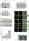
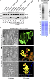
Binding of Vps4 to ESCRT-III is mainly mediated by Snf7 and Vps2. (A) The schematic representation shows ESCRT-III, the Snf7 homooligomer (blue dots), the MIM2 of Vps20 (green square), the MIM2 of Snf7 (blue square), the MIM1 of Vps24 (violet circle), the MIM1 of Vps2 (red circle), and its interaction with the MIT domain (green) of Vps4. N, N terminus; C, C terminus. (B, C, E, and F) Experiments were analyzed by SDS-PAGE and Western blotting. (B) Total cell lysates from WT cells and the vps20*, snf7*, vps24*, vps2* quadruple mutant. (C) Immunoprecipitation (IP) of Vps4-HA from cell lysates of WT cells and the indicated mutants. #, unspecific signal. (D) Quantification (MaxQuant) of mixed Vps4-HA immunoprecipitates from WT cells (labeled with [13C6/15N2] l-lysine) and from vps2* mutants. Heavy (H) to light (L) ratios of the ESCRT-III proteins and Vps4. Heavy/light = 1, equal peptides from WT (heavy) and vps2* (light); heavy/light > 1, more peptides from the WT (heavy). (E) Membrane fraction (M) and cytoplasmic fraction (C) of WT cells and the indicated mutants expressing Vps4-E233Q. (F) Immunoprecipitation of Vps4-E233Q-HA from cell lysates of the indicated MIM mutants. IN, input; n.a., not annotated.
Figure 2.
Figure 2.
The interaction of Vps4 with Snf7 and Vps2 is essential for efficient ESCRT-III disassembly and MVB sorting. (A–C) Experiments were analyzed by SDS-PAGE and Western blotting. (A) Solubilized membrane fractions (13,000 g pellet) of WT cells and the indicated MIM mutants were subjected to velocity sedimentation. (B) Membrane fractions (M) and cytoplasmic fractions (C) of WT cells and the indicated mutants. (C) Semi–in vitro disassembly assay with membrane fractions isolated from vps4Δ mutants in combination with the indicated MIM mutations. Membrane fractions were incubated with ATP and 100 nM of recombinant Vps4 for 0, 30, or 60 s. Membrane-associated proteins (13,000 g pellet [P]) and released proteins (13,000 g supernatant [S]) were separated by centrifugation. (D) Live-cell fluorescence microscopy of WT cells and the indicated mutants expressing GFP-CPS, FM4-64, vacuole (V), and the class E compartment (E). Bar, 5 µm. Schematic presentation of GFP-CPS–sorting phenotypes. DIC, differential interference contrast. (E) GFP-CPS–sorting phenotypes of all ESCRT-III MIM mutant combinations. (F) Quantification of the LUCID assay in WT cells and the indicated mutants. Ratios of the Sna3-FLuc reporter activity to the cytoplasmic Renilla luciferase (RLuc) activity normalized with SDs are shown; n = 6. (G) Dilution series of WT cells and the indicated mutants were grown on yeast nitrogen base plates at 26 or 37°C. **, P < 0.01; ***, P < 0.001.
Figure 3.
Figure 3.
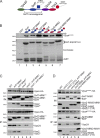
Rearranging the binding of Vps4 to the ESCRT-III complex. (A) Schematic representation of the ESCRT-III complex. (B) In vitro pull-down assay with ESCRT-III chimeras. Their domain organization is shown. GST alone or the indicated GST–ESCRT-III chimeras were immobilized on beads and incubated for 10 min at RT with Vps4-E233Q and 1 mM ATP. Bound fractions were analyzed by SDS-PAGE, Western blotting (WB), or Coomassie staining. (C and D) Vps4-HA (C) or Vps4-E233Q-HA (D) were immunoprecipitated (IP) from cell lysates of the indicated mutants and analyzed by SDS-PAGE and Western blotting. IN, input.
Figure 4.
Figure 4.
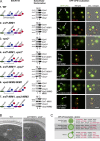
The MIMs of Vps2 and Snf7 couple ESCRT-III disassembly to ILV biogenesis. (A) Sections 1–7 show schematic representation of ESCRT-III complexes, the corresponding subcellular fractionation, and live-cell microscopy using GFP-CPS and FM4-64. Bar, 5 µM. Membrane fractions (M) and cytoplasmic fraction (C) of the indicated mutants were analyzed by SDS-PAGE and Western blotting. The uncut films are shown in Fig. S3 A. V, vacuole; E, class E compartment; DIC, differential interference contrast. (B) Transmission EM of cryofixed WT cells and snf7-MIM1, vps2* mutants with class E compartments. Bar, 250 nm. (C) GFP-CPS–sorting phenotypes of the tested strains with different chimeric ESCRT-III complexes.
Figure 5.
Figure 5.
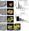
Vps4-mediated ESCRT-III disassembly controls ILV biogenesis in vivo. (A) Electron tomography of cryofixed WT cells overexpressing Vps21 (TDH3-VPS21) and the indicated mutants. 2D slices from tomographic reconstructions and models from 400-nm sections are shown. Limiting MVB membrane (yellow), ILVs (red), nuclear envelope (blue), and class E–like structures (green) are shown. Bars, 150 nm. Arrowheads point to enlarged budding profiles in the snf7*, vps2* double mutants. (B) Size distribution (in 10-nm steps) of ILV diameters in WT cells and the indicated mutants. (C) Mean number of ILVs/MVB for ≥10 fully reconstructed MVBs from WT cells and the indicated mutants. Error bars indicate the SDs. *, P < 0.05; ***, P < 0.001.
Figure 6.
Figure 6.
Slow and continuous ESCRT-III recycling and ILV biogenesis in snf7*, vps2* double mutants. (A) vps4-ts mutants, vps4Δ mutants and snf7*, vps2*, vps4-ts mutants were shifted to the nonpermissive temperature (37°C) for 4 h. 15 min before cells were shifted back to 26°C, 50 µg/ml cycloheximide was added. Cells were incubated for 2 and 4 h at 26°C. Subcellular fractionation was performed at the indicated time points. Cytoplasmic fractions were analyzed by SDS-PAGE and Western blotting. (B) Cells were incubated with cycloheximide (CHX) and labeled for 5 min with [35S]methionine. Autoradiogram and Coomassie staining of cell lysates is shown. (C) Electron tomography of cryofixed snf7*, vps2*, vps4-ts mutants at the indicated temperatures and times. 2D slices from tomographic reconstructions and models from 400-nm sections are shown. Arrowheads point to enlarged budding profiles. Limiting MVB membrane (yellow), ILVs (red), vacuolar membrane (brown), nuclear envelope (blue), and class E–like structures (green) are depicted. Bars, 150 nm.
Figure 7.
Figure 7.
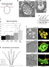
Binding of Vps4 to Snf7 and Vps2 is required for ILV neck constriction. (A) Schematic representation of ILV budding profiles as detected in tomographic reconstructions. (B) MVB of WT cells and MVB-like structure from snf7*, vps2* double mutants. The arrows indicate ILV necks. (C) Mean membrane neck diameters measured in the tomograms. Error bars indicate the SD of the membrane neck diameters measured in the tomograms. (D) Size distribution of individual membrane neck diameters of the WT and the indicated mutants. (E) Membrane fraction (M) and cytoplasmic fraction (C) of the indicated mutants were analyzed by SDS-PAGE and Western blotting. (F) 2D slices and 3D models from 400-nm sections of the indicated mutants overexpressing Vps21 (TDH3-VPS21). Limiting MVB membrane (yellow), ILVs (red), vacuolar membrane (brown), and class E compartments (green) are shown. Bars, 150 nm. ***, P < 0.001.
Figure 8.
Figure 8.
Proposed model for ILV biogenesis. PI(3)P, phosphatidylinositol 3-phosphate.

References

    1. Adell M.A., Teis D. 2011. Assembly and disassembly of the ESCRT-III membrane scission complex. FEBS Lett. 585:3191–3196 10.1016/j.febslet.2011.09.001 - DOI - PMC - PubMed
    1. Azmi I., Davies B., Dimaano C., Payne J., Eckert D., Babst M., Katzmann D.J. 2006. Recycling of ESCRTs by the AAA-ATPase Vps4 is regulated by a conserved VSL region in Vta1. J. Cell Biol. 172:705–717 10.1083/jcb.200508166 - DOI - PMC - PubMed
    1. Azmi I.F., Davies B.A., Xiao J., Babst M., Xu Z., Katzmann D.J. 2008. ESCRT-III family members stimulate Vps4 ATPase activity directly or via Vta1. Dev. Cell. 14:50–61 10.1016/j.devcel.2007.10.021 - DOI - PubMed
    1. Babst M., Sato T.K., Banta L.M., Emr S.D. 1997. Endosomal transport function in yeast requires a novel AAA-type ATPase, Vps4p. EMBO J. 16:1820–1831 10.1093/emboj/16.8.1820 - DOI - PMC - PubMed
    1. Babst M., Wendland B., Estepa E.J., Emr S.D. 1998. The Vps4p AAA ATPase regulates membrane association of a Vps protein complex required for normal endosome function. EMBO J. 17:2982–2993 10.1093/emboj/17.11.2982 - DOI - PMC - PubMed

Publication types

MeSH terms

Substances