. 2014 Apr 14;205(1):51-65.
doi: 10.1083/jcb.201311057. Epub 2014 Apr 7.

Substrate-gated docking of pore subunit Tha4 in the TatC cavity initiates Tat translocase assembly

Affiliations

Substrate-gated docking of pore subunit Tha4 in the TatC cavity initiates Tat translocase assembly

Cassie Aldridge et al. J Cell Biol. .

Abstract

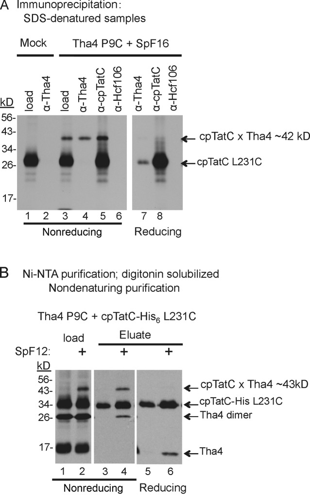
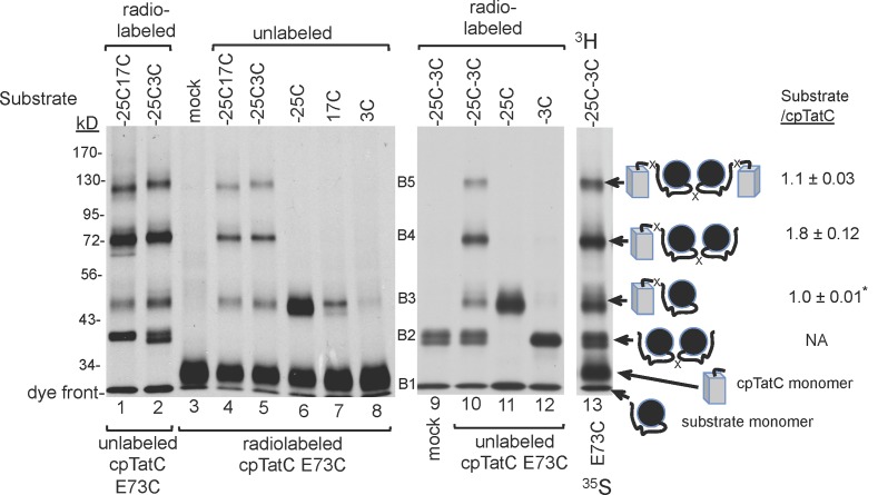
The twin-arginine translocase (Tat) transports folded proteins across tightly sealed membranes. cpTatC is the core component of the thylakoid translocase and coordinates transport through interactions with the substrate signal peptide and other Tat components, notably the Tha4 pore-forming component. Here, Cys-Cys matching mapped Tha4 contact sites on cpTatC and assessed the role of signal peptide binding on Tha4 assembly with the cpTatC-Hcf106 receptor complex. Tha4 made contact with a peripheral cpTatC site in nonstimulated membranes. In the translocase, Tha4 made an additional contact within the cup-shaped cavity of cpTatC that likely seeds Tha4 polymerization to form the pore. Substrate binding triggers assembly of Tha4 onto the interior site. We provide evidence that the substrate signal peptide inserts between cpTatC subunits arranged in a manner that conceivably forms an enclosed chamber. The location of the inserted signal peptide and the Tha4-cpTatC contact data suggest a model for signal peptide-gated Tha4 entry into the chamber to form the translocase.


Figures

Figure 1.
Figure 1.
Contact between the Tha4 TM and cpTatC TM4 in the translocase. (A) Radiolabeled pre-cpTatC L231C was imported into chloroplasts. Recovered thylakoids were incubated with in vitro–translated unlabeled Tha4 single Cys variants (XnC) followed by in vitro–translated SpF16 Tat substrate (see Materials and methods). Samples at 15°C were illuminated to assemble the translocase and CuP was added to promote disulfide formation (Materials and methods). Analysis was by SDS-PAGE/fluorography under nonreducing or reducing (+ β-mercaptoethanol) conditions. (B) Assays received SpF16 or mock translation extract before cross-linking. (C) Assays received either the full-size substrate tOE17-20F or the inactive twin lysine variant (KK-tOE17-20F). (D) Models of Tha4 and cpTatC with the positions of Cys substitutions marked with stars. Tha4 F4C E10Q is a nonfunctional Tha4 variant.
Figure 2.
Figure 2.
The ∼42-kD band is a cross-linking product between Tha4 and cpTatC. (A) Thylakoids with radiolabeled cpTatC L231C were incubated with mock translation extract or unlabeled Tha4 P9C followed by mock translation extract or SpF16. After disulfide cross-linking, recovered thylakoids were subjected to denaturing immunoprecipitation with antibody-linked beads as shown above the panels (see Materials and methods). (B) Thylakoids containing radiolabeled cpTatC–His6 L231C were incubated with radiolabeled Tha4 P9C and either SpF12 or mock translation extract and then subjected to disulfide cross-linking. Recovered thylakoids were dissolved in 1% digitonin and cpTatC-His6 purified on magnetic Ni-NTA beads (Materials and methods). Eluted samples in A and B were analyzed by nonreducing or reducing SDS-PAGE/fluorography as in Fig. 1.
Figure 3.
Figure 3.
Stromal domains of cpTatC contact the stromal domain of Tha4; lumenal domains of cpTatC contact the lumen-proximal domain of Tha4. Thylakoid membranes containing radiolabeled cpTatC single Cys variants were incubated with unlabeled Tha4 single Cys variants and SpF16 or with mock translation extract (−) as in Fig. 1 and subjected to disulfide cross-linking (see Materials and methods). Samples were analyzed by nonreducing SDS-PAGE/fluorography. (A) cpTatC with Cys residues in transmembrane domains. (B) cpTatC with Cys residues in stroma-exposed and lumen-exposed domains.
Figure 4.
Figure 4.
Tha4 contacts lumenal loop L3 of cpTatC constitutively, and TM4 and TM5 in a substrate-enhanced manner. (A and B) Thylakoid membranes containing radiolabeled cpTatC Cys received unlabeled Tha4 Cys variants and either mock translation extract (−) or SpF16, and were subjected to disulfide cross-linking (see Materials and methods). (C) The effects of substrate binding and translocase assembly on cpTatC dimerization directed by lumen proximal L126C, V270C, and T275C. Thylakoids with imported radiolabeled cpTatC-Cys were incubated with mock translation extract, unlabeled SpF16 plus mock translation extract, and SpF16 plus unlabeled wild-type Tha4 as shown. Disulfide cross-linking was as in Fig. 1 and BMOE cross-linking as in the Materials and methods, except that only the reactions with Tha4 plus SpF16 received light during the reaction and pre-incubation.
Figure 5.
Figure 5.
Substrate-enhanced contacts between the Tha4 TM and cpTatC TM4 and TM5 also require the proton gradient. Thylakoid membranes containing radiolabeled cpTatC Cys variants in TM4 (L231C), TM5 (Y259C or V270C), or L3 (T275C) were incubated with unlabeled Tha4 Cys variants and mock translation extract or SpF12, and were subjected to disulfide cross-linking. All reactions were preincubated in light during Tha4 integration (see Materials and methods). Incubations for cross-linking in A were in darkness. Incubations in B and D were in the light. As designated by Nig/Val +, certain reactions received 1 µM nigericin and 2 µM valinomycin immediately after SpF12 addition to dissipate the proton gradient. (C) The cpTatC x Tha4 cross-linking products in A and B were quantified by densitometric analysis of films scanned by light transmission and analyzed with ImageJ software. The averages and differences from the mean were from two repeats of the experiment. All values from each film were normalized to the density of the cpTatC V270C × Tha4 P9C plus SpF12 band, which was assigned an arbitrary value of 100.
Figure 6.
Figure 6.
The H-domain of the substrate signal peptide makes contact with the cpTatC concave surface at TM5. Thylakoid membranes containing radiolabeled cpTatC Cys variants were incubated in a binding reaction with tOE17-20F substrate Cys variants as shown above the panels. After washing, thylakoids were subjected to disulfide cross-linking at 15°C. (A) Diagram of the signal peptide and early mature domain of the substrate showing numbering convention. The H-domain is underlined. (B) Survey of cpTatC Cys with substrate containing Cys in the signal peptide or early mature domain. (C, left) Demonstration that the ∼48-kD band is a cross-linking product between substrate −7C and cpTatC V270C. Cross-linking reactions were conducted in which substrate or cpTatC-Cys was absent, or in which only one or both were radiolabeled as shown above the panel. (Right) The cpTatC TM5 V270C was combined with substrate containing a range of single Cys substitutions. The ∼46-kD band produced from substrate −10C (asterisk) is a nonspecific cross-linking product.
Figure 7.
Figure 7.
Substrate protein signal peptides insert between cpTatC subunits. Thylakoid membranes containing radiolabeled or unlabeled cpTatC E73C (domain S1, shown below the panels) were incubated in a binding reaction with radiolabeled or unlabeled single and double Cys variants of the full-length tOE17-20F substrate as shown above the panels. Disulfide cross-linking was conducted as described in the Materials and methods. The identification of B3 as substrate × cpTatC is inferred from the migration of the cross-linking product between cpTatC E73C and substrate −25C (lanes 6 and 11). The identification of band B2 as substrate dimer is inferred from the migration of the bands from the reaction containing radiolabeled substrate −3C (lane 12). Identification of the B4 band as substrate2 × cpTatC and the B5 band as substrate2 × cpTatC2 was based on their Mr and the substrate/cpTatC abundance in the band. The ratio of abundance was determined in an experiment in which substrate was labeled with 3H-leucine and cpTatC was labeled with 35S-methionine (lane 13). The ratio of tritium to 35S for the B3 band was set as 1:1 (asterisk) and the ratios of B4 and B5 calculated normalized to that value. Ratios are the average and standard deviation obtained from six individual cross-linking assays.
Figure 8.
Figure 8.
Models for the association of cpTatC, Tha4, and substrate during assembly of the Tat translocase. (A) A cartoon model for pea chloroplast TatC structure based on homology with the A. aeolicus TatC structure (see Materials and methods) with all Cys substitutions tested in this study colored yellow on the cpTatC backbone and labeled by residue number; yellow highlighted residue numbers produced disulfide cross-links with Tha4. Residues that, when mutated to alanine, impair twin arginine binding are colored orange. Glutamine 234, proposed to mediate docking of Tha4 E10, is colored magenta. The carboxyl-proximal helix of cpTatC TM5 is colored cyan. (B) Proposed arrangement of cpTatC, bound substrates, and the TMs of Tha4 (red cylinders) and Hcf106 (green cylinders) in a dimer of cpTatC. A ribbon depiction of the proposed cpTatC dimer as viewed from the chloroplast stroma with cpTatC subunits colored blue and purple, respectively, is shown. The suggested substrate arrangement is shown in yellow with the signal peptide hydrophobic helix associated with the Hcf106 TM and the carboxyl-proximal signal peptide and N-terminal residues of the substrate mature domain in the central cavity. A stippled yellow line depicts the segment that directs disulfide-linked substrate dimers. (C and D) Interpretation of the positions of Tha4 and Hcf106 TMs with cpTatC constitutively (C) and in the translocase (D) are based on disulfide cross-linking and the position of the E. coli TatB (Hcf106 orthologue; Rollauer et al., 2012). The N-terminal segments of Tha4 and Hcf106 were modeled onto the E. coli TatA structure (see Materials and methods).

Similar articles

Cited by

References

    1. Alami M., Lüke I., Deitermann S., Eisner G., Koch H.G., Brunner J., Müller M. 2003. Differential interactions between a twin-arginine signal peptide and its translocase in Escherichia coli. Mol. Cell. 12:937–946 10.1016/S1097-2765(03)00398-8 - DOI - PubMed
    1. Alcock F., Baker M.A., Greene N.P., Palmer T., Wallace M.I., Berks B.C. 2013. Live cell imaging shows reversible assembly of the TatA component of the twin-arginine protein transport system. Proc. Natl. Acad. Sci. USA. 110:E3650–E3659 10.1073/pnas.1306738110 - DOI - PMC - PubMed
    1. Alder N.N., Theg S.M. 2003. Energetics of protein transport across biological membranes. a study of the thylakoid DeltapH-dependent/cpTat pathway. Cell. 112:231–242 10.1016/S0092-8674(03)00032-1 - DOI - PubMed
    1. Aldridge C., Storm A., Cline K., Dabney-Smith C. 2012. The chloroplast twin arginine transport (Tat) component, Tha4, undergoes conformational changes leading to Tat protein transport. J. Biol. Chem. 287:34752–34763 10.1074/jbc.M112.385666 - DOI - PMC - PubMed
    1. Arnon D.I. 1949. Copper enzymes in isolated chloroplasts. Polyphenol oxidase in Beta vulgaris. Plant Physiol. 24:1–15 10.1104/pp.24.1.1 - DOI - PMC - PubMed

Publication types

MeSH terms

LinkOut - more resources