. 2014 Apr 14;205(1):113-26.
doi: 10.1083/jcb.201312072. Epub 2014 Apr 7.

A novel probe for phosphatidylinositol 4-phosphate reveals multiple pools beyond the Golgi

Affiliations

A novel probe for phosphatidylinositol 4-phosphate reveals multiple pools beyond the Golgi

Gerald R V Hammond et al. J Cell Biol. .

Abstract

Polyphosphoinositides are an important class of lipid that recruit specific effector proteins to organelle membranes. One member, phosphatidylinositol 4-phosphate (PtdIns4P) has been localized to Golgi membranes based on the distribution of lipid binding modules from PtdIns4P effector proteins. However, these probes may be biased by additional interactions with other Golgi-specific determinants. In this paper, we derive a new PtdIns4P biosensor using the PtdIns4P binding of SidM (P4M) domain of the secreted effector protein SidM from the bacterial pathogen Legionella pneumophila. PtdIns4P was necessary and sufficient for localization of P4M, which revealed pools of the lipid associated not only with the Golgi but also with the plasma membrane and Rab7-positive late endosomes/lysosomes. PtdIns4P distribution was determined by the localization and activities of both its anabolic and catabolic enzymes. Therefore, P4M reports a wider cellular distribution of PtdIns4P than previous probes and therefore will be valuable for dissecting the biological functions of PtdIns4P in its assorted membrane compartments.


Figures

Figure 1.
Figure 1.
Localization of fluorescent protein fusions of the P4M domain (residues 546–647) from L. pneumophila SidM/DrrA protein. (A) GFP-P4M expressed in COS-7 cells and imaged on a spinning-disk confocal; shown are single confocal optical sections along the indicated axes, a 3D maximum intensity projection of 38 optical sections acquired at 0.2-µm intervals, or a color-coded temporal (3Dt) projection of 23 maximum intensity projections acquired at 8.15-s intervals. (B) Colocalization of the iRFP-tagged P4M domain with the PH domains from PLCδ1 and FAPP1. The color-coded images are normalized mean deviation product (nMDP) images of the indicated image pairs, showing the spatial distribution of colocalized pixels. mCh, mCherry. (C) Two P4M domains fused in tandem to GFP are shown as confocal optical sections along the indicated axes. Note the collapsed Golgi morphology. Bars, 10 µm.
Figure 2.
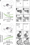
Figure 2.
P4M domain localization is PtdIns4P dependent at the Golgi, which contains low levels of PtdIns(4,5)P2. (A) Rapamycin (rapa)-induced recruitment of PJ-Sac to the FRB-tagged tail domain (residues 3,140–3,269) from human giantin (GOLGB1) induces dephosphorylation of PtdIns4P (PI4P) to PtdIns (PI), releasing GFP-P4M from the Golgi. No change is induced by the catalytically inactive PJ-Dead construct. Graphs show normalized intensity of GFP-P4M or PH-PLCδ1-GFP at the Golgi (defined by the FRB-giantintail mask) after treatment with 1 µM rapamycin. Data are grand means ± SEM of eight independent experiments. Significant (P < 0.05) changes are denoted as the largest p-value over the indicated range (two-way ANOVA; see Materials and methods). Images show representative cells with the Golgi localization of the FRB-mCherry-giantintail recruiter and its conversion to a binary mask for quantification of GFP-P4M fluorescence. Also shown is the localization of mRFP-tagged PJ-Sac and GFP-P4M before and after addition of 1 µM rapamycin. (B) Rapamycin-induced recruitment of FKBP-PIP5K to FRB-giantintail stimulates phosphorylation of PtdIns4P to produce PtdIns(4,5)P2 (PI(4,5)P2), recruiting PH-PLCδ1-GFP. No effect is observed for the catalytically inactive D253A mutant. Data are grand means of three or four independent experiments ± SEM. Insets depict an enlarged view of the regions depicted by hashed boxes. Bars: (main images) 10 µm; (insets) 5 µm.
Figure 3.
Figure 3.
P4M Golgi localization is independent of Arf1. (A) COS-7 cells expressing mCherry-P4M and PH-FAPP1-GFP before and 20 min after the addition of 5 µg/ml Brefeldin A (BFA). (B) Normalized intensity of PH-FAPP1-GFP and mCherry (mCh)-P4M at the Golgi after treatment with 5 µM Brefeldin A. Data are grand means ± SEM of three independent experiments. Significant (P < 0.05) changes are denoted as the largest p-value over the indicated range (two-way ANOVA; see Materials and methods). (C) COS-7 cells expressing GFP-P4M and ManII1–102–mKusabira Orange before and 20 min after the addition of 5 µg/ml Brefeldin A. (D) Normalized intensity of GFP-P4M and ManII1–102–mKusabira Orange; data are means ± SEM of seven cells from two independent experiments. Bars, 10 µm.
Figure 4.
Figure 4.
P4M localization at the PM depends on PtdIns4P. (A) Rapamycin (rapa)-induced recruitment of PJ-Sac to the PM via dimerization with FRB targeted to the PM causes depletion of PtdIns4P (PI4P) and release of GFP-P4M from the membrane, without effect on the PtdIns(4,5)P2 (PI(4,5)P2) probe iRFP-PH-PLCδ1. (B) Recruitment of an INPP5E domain to the PM causes breakdown of PtdIns(4,5)P2 into PtdIns4P, causing release of iRFP-PH-PLCδ1 and increased PM binding of GFP-P4M. (C) PM recruitment of PJ (containing both Sac and INPP5E domains) causes breakdown of PtdIns(4,5)P2 and PtdIns4P into PtdIns, resulting in release of both probes from the PM. (D) No effect on either probe is seen when catalytically inactive mutations in the Sac and INPP5E domains are introduced into PJ. (E and F) PM intensity of cells expressing either PtdIns4P-binding GFP-P4M or GFP-PH-OSBP during recruitment of PJ-Sac (E) or INPP5E (F) was measured by TIRF. Images show the FRB domain fused to CFP and the PM-targeted acylated Lyn11 peptide and the resulting binary masks used to measure PM intensity as well as the GFP-P4M and iRFP PH-PLCδ1 domains before and after enzyme recruitment with rapamycin. Graphs are from three (E) or four (A–D and F) independent experiments and are grand means ± SEM. Statistical significance (P < 0.05) is depicted over the indicated range (two-way ANOVA; see Materials and methods). n.s. = i.e., P > 0.05. Bars, 10 µm.
Figure 5.
Figure 5.
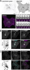
P4M binds endosomes/lysosomes. (A) COS-7 cells expressing mCherry (mCh)-tagged P4M were loaded with 100 nM LysoTracker green. The insets 1 and 2 are shown as a montage of images acquired at 3.9-s intervals showing motile, P4M, and LysoTracker-positive structures highlighted by the arrowheads. (B) COS-7 cells transfected with GFP-P4M, the late endosomal marker Rab7 (tagged with mTurquoise2 [mTq2]), and the early endosomal marker Rab5 (tagged with mCherry). A binary mask derived from the Rab7 and Rab5 images was used to calculate the relative intensity of the P4M signal associated with each compartment. The results from 24 cells are shown as a paired scatter plot. (C) As in B, except cells were transfected with the GFP-tagged FYVE domain from EEA1 as a marker of PtdIns3P. Bars: (main images) 10 µm; (insets) 5 µm. Statistics report the p-value from a paired Student’s t test (see Materials and methods).
Figure 6.
Figure 6.
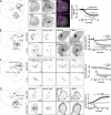
P4M localization is dependent on endosomes/lysosomal PtdIns4P. (A) COS-7 cells transfected with the PtdIns3P (PI3P) reporter GFP-FYVE-EEA1 and mCherry-P4M imaged by spinning-disk confocal microscopy during treatment with 100 nM wortmannin, a PtdIns 3-kinase (PI3K) inhibitor. (B) FRB targeted to late endosomes/lysosomes by fusion to Rab7 recruits lipid phosphatase PJ-Sac (active against PtdIns4P, PtdIns3P, and PtdIns(3,5)P2) or MTM1 (active against PtdIns3P and PtdIns(3,5)P2 but not PtdIns4P) in cells transfected with GFP-P4M. (C) FRB targeted to early endosomes by fusion to Rab5 recruits PJ-Sac or MTM1 in cells transfected with GFP-FYVE-EEA1. (D) FRB-Rab7 recruits a PIP5K or its catalytically inactive D253A mutant; the effect on PtdIns(4,5)P2 generation is monitored with the PtdIns(4,5)P2 reporter PH-PLCδ1-GFP. Graphs are grand means ± SEM from three independent experiments. Statistical significance (P < 0.05) is depicted over the indicated range (two-way ANOVA; see Materials and methods). n.s. = i.e., P > 0.05. rapa, rapamycin. Bars, 10 µm.
Figure 7.
Figure 7.
GFP-P4M detects ectopic PtdIns4P synthesis in the ER driven by hepatitis C nonstructural protein 5A. COS-7 cells transfected with GFP-P4M, NS5A-mCherry, and mTq2-tagged PI4KA wild type (WT) or its catalytically inactive D1957A mutant. Colocalization between GFP-P4M and NS5A-mCherry (mCh) is depicted in the nMDP images. The box and whisker plot shows the nMDP values for 30 cells (boxes denote the interquartile range with lines at the median; whiskers denote the 10th and 90th percentiles). The p-value was generated by a Mann–Whitney U test. Bar, 10 µm.
Figure 8.
Figure 8.
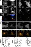
GFP-P4M reveals pools of PtdIns4P accessible to all of the lipid’s anabolic and catabolic enzymes. (A) COS-7 cells transfected with mCherry (mCh)-P4M and a fluorescent protein conjugate of each of the four human PI4K isoforms. Cells expressing mTq2-tagged PI4KA and mCherry-P4M were cotransfected with GFP-EFR3B and iRFP-TTC7B (insets) that together recruit the kinase to the PM. Colocalizing pixels are highlighted in the nMDP imaged. (B) Cells transfected with GFP-P4M and mCherry-tagged Sac proteins: either wild type (WT), the C389S catalytically inactive mutant, leucine-zipper alanine (LZA) ER export–deficient mutant, or K2A Golgi retrieval–deficient mutant. The graphs show the relative P4M intensity at the Golgi, PM, and Rab7-positive compartments (box and whiskers as in Fig. 7; boxes denote the interquartile range with lines at the median; whiskers denote the 10th and 90th percentiles). The numbers above each group refer to the p-value (one-way ANOVA, 30 cells per group; see Materials and methods) compared with the C389S mutant or as indicated. Bars, 10 µm.

Similar articles

Cited by

References

    1. Baird D., Stefan C., Audhya A., Weys S., Emr S.D. 2008. Assembly of the PtdIns 4-kinase Stt4 complex at the plasma membrane requires Ypp1 and Efr3. J. Cell Biol. 183:1061–1074 10.1083/jcb.200804003 - DOI - PMC - PubMed
    1. Balla A., Tuymetova G., Barshishat M., Geiszt M., Balla T. 2002. Characterization of type II phosphatidylinositol 4-kinase isoforms reveals association of the enzymes with endosomal vesicular compartments. J. Biol. Chem. 277:20041–20050 10.1074/jbc.M111807200 - DOI - PubMed
    1. Balla A., Tuymetova G., Tsiomenko A., Várnai P., Balla T. 2005. A plasma membrane pool of phosphatidylinositol 4-phosphate is generated by phosphatidylinositol 4-kinase type-III alpha: studies with the PH domains of the oxysterol binding protein and FAPP1. Mol. Biol. Cell. 16:1282–1295 10.1091/mbc.E04-07-0578 - DOI - PMC - PubMed
    1. Balla A., Tuymetova G., Tóth B., Szentpetery Z., Zhao X., Knight Z.A., Shokat K., Steinbach P.J., Balla T. 2008a. Design of drug-resistant alleles of type-III phosphatidylinositol 4-kinases using mutagenesis and molecular modeling. Biochemistry. 47:1599–1607 10.1021/bi7017927 - DOI - PubMed
    1. Balla A., Kim Y.J., Várnai P., Szentpetery Z., Knight Z., Shokat K.M., Balla T. 2008b. Maintenance of hormone-sensitive phosphoinositide pools in the plasma membrane requires phosphatidylinositol 4-kinase IIIalpha. Mol. Biol. Cell. 19:711–721 10.1091/mbc.E07-07-0713 - DOI - PMC - PubMed

Publication types

MeSH terms

Substances