. 2014 Jul;82(7):2688-96.
doi: 10.1128/IAI.00055-14. Epub 2014 Apr 7.

Lack of long-lasting hydrosalpinx in A/J mice correlates with rapid but transient chlamydial ascension and neutrophil recruitment in the oviduct following intravaginal inoculation with Chlamydia muridarum

Affiliations

Lack of long-lasting hydrosalpinx in A/J mice correlates with rapid but transient chlamydial ascension and neutrophil recruitment in the oviduct following intravaginal inoculation with Chlamydia muridarum

Hongbo Zhang et al. Infect Immun. 2014 Jul.

Abstract

Lower genital tract infection with Chlamydia trachomatis and C. muridarum can induce long-lasting hydrosalpinx in the upper genital tract of women and female mice, respectively. However, A/J mice were highly resistant to induction of long-lasting hydrosalpinx by C. muridarum. We further compared host inflammatory responses and chlamydial infection courses between the hydrosalpinx-resistant A/J mice and CBA/J mice known to be susceptible to hydrosalpinx induction. Both mouse strains developed robust pyosalpinx during the acute phase followed by hydrosalpinx during the chronic phase. However, the hydrosalpinges disappeared in A/J mice by day 60 after infection, suggesting that some early hydrosalpinges are reversible. Although the overall inflammatory responses were indistinguishable between CBA/J and A/J mice, we found significantly more neutrophils in oviduct lumen of A/J mice on days 7 and 10, which correlated with a rapid but transient oviduct invasion by C. muridarum with a peak infection on day 7. In contrast, CBA/J mice developed a delayed and extensive oviduct infection. These comparisons have revealed an important role of the interactions of oviduct infection with inflammatory responses in chlamydial induction of long-lasting hydrosalpinx, suggesting that a rapid but transient invasion of oviduct by chlamydial organisms can prevent the development of the long-lasting hydrosalpinges.


Figures

FIG 1
FIG 1
Development of long-lasting hydrosalpinx in CBA/J but not A/J mice following C. muridarum infection. Both CBA/J (n = 10) and A/J (n = 10) mice were intravaginally infected with 2 × 10⁵ IFUs of C. muridarum, and 60 days after infection all mice were sacrificed for observing hydrosalpinx. (A) Images of whole genital tracts from all 20 mice are presented in the left columns (a to j for CBA/J, k to t for A/J) with vagina on the left and oviduct/ovary on the right sides. The images for areas covering the oviduct/ovary portions were magnified and are shown to the right of the corresponding whole-genital-tract images, with hydrosalpinx marked with red arrows and hydrosalpinx severity scores indicated in white numbers. (B) The hydrosalpinx severity scores and incidence rates (data not shown) are listed along the y axis. Mice with hydrosalpinx in either or both oviducts were considered positive for hydrosalpinx. The severity of both hydrosalpinges from a given mouse was scored separately and added together as the severity score assigned to the particular mouse. Note that A/J mice failed to develop any significant hydrosalpinx, with both hydrosalpinx incidence (a, Fisher's exact test) and severity (b, Wilcoxon rank-sum test) significantly lower than those of CBA/J mice (**, P < 0.01 for both).
FIG 2
FIG 2
Monitoring oviduct gross pathology along C. muridarum infection course. CBA/J and A/J mice were infected with C. muridarum as described in the legend to Fig. 1 and sacrificed for visual observation of oviduct pathology, including both pyosalpinx and hydrosalpinx, on days 3 (n = 5 for CBA/J and A/J groups, respectively), 7 (n = 5), 10 (n = 6), 14 (n = 5), 21 (n = 5), 28 (n = 5), 35 (n = 5), and 60 (n = 10) after inoculation. One or two representative images of oviduct/ovary from both sides of a mouse genital tract are presented for both strains of mice at each time point, except for days 3 and 7 (no gross pathology could be detected on these early days). The visually observable pyosalpinx (Pyo) was marked with yellow and hydrosalpinx (Hydro) with red arrows, while the gross pathology severity was scored according to the criteria described in Materials and Methods and marked with numbers in white in corresponding images. To validate the visually observable gross pathology, H&E-stained sections were prepared from the corresponding genital tract tissues (Histopath.). Representative images from each strain of mice, taken under a 10× and 100× objective lens, respectively, are shown with pyosalpinx, hydrosalpinx, or normal oviducts marked with P (in yellow), H (red), or N (blue), respectively. White rectangles in images of a 10× objective lens indicate the areas where the images were further magnified under the 100× objective lens. Note that oviducts with pyosalpinx (yellow P) were filled with inflammatory infiltrates, while those with hydrosalpinx (red H) showed empty lumenal space suggestive of clear solution accumulation. Inflammation was also observed under microscopy in oviducts without visually detectable pathology.
FIG 3
FIG 3
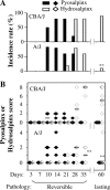
Scoring oviduct gross pathology along C. muridarum infection course. Both the gross pathology incidence rates (A) and severity scores (B) were calculated from each group of mice presented in Fig. 2. The values are listed along the y axis. Note that extensive pyosalpinx (solid bars or diamonds) was observed in both strains of mice during the first 4 weeks after infection followed by or overlapping hydrosalpinx (open bars or diamonds) in both strains of mice. However, by day 60, hydrosalpinx incidence was significantly higher in CBA/J mice (*, P < 0.01 by Fisher's exact; also see the legend to Fig. 1), and by days 35 and 60, hydrosalpinx severity was significantly lower in A/J mice (*, P < 0.01 by Wilcoxon rank-sum test).
FIG 4
FIG 4
Monitoring oviduct inflammatory histopathology following C. muridarum infection. The same oviduct tissues from the experiments described for Fig. 2 were subjected to H&E staining for evaluating inflammatory histopathology. (A) Representative images from each group, taken under 10× (left column, a to g in CBA/J mice and h to n in A/J mice) and 100× objective lenses (right column, a1 to g1 and a2 to g2 in CBA/1J mice and h1 to n1 and h2 to n2 in A/J mice) are shown. White rectangles in images from the 10× objective lens indicate the same areas from which the right-side images were taken under a 100× objective lens. (B) The severity of inflammatory infiltration or exudation in oviduct tissue (bottom) and lumen (top) areas was scored separately as described in Materials and Methods and is listed along the y axis (solid diamonds for CBA/J, open diamonds for A/J). Note that both groups of mice developed extensive inflammatory histopathology that peaked on day 14 after infection in both oviduct tissue and lumen (*, P < 0.05 by Wilcoxon rank-sum test).
FIG 5
FIG 5
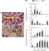
Detecting oviduct inflammatory cells following C. muridarum infection. The H&E-stained sections described in the legend to Fig. 4 were reexamined for identifying neutrophils and mononuclear cells. (A) Example images marked with either neutrophils (red arrows) or mononuclear cells (yellow arrows) are shown. In the image taken from an oviduct tissue section with an acute infection, more neutrophils were detected (a, A/J mice), while at the chronic stage, more mononuclear inflammatory cells, including plasma cells, lymphocytes, and macrophages, were detected (b, CBA/J mice). Total inflammatory cells and neutrophil and mononuclear cell subpopulations were counted in individual 100× objective lens views (for high-density infiltration) or the entire oviduct tissue or lumen section (for scarce infiltration). (B) The number of neutrophils (a and b) and mononuclear cells (c and d) per view from either oviduct lumen (a and c) or tissue (b and d) areas of each mouse was used to calculate means and standard deviations for each group as shown along the y axis. Note that both CBA/J (solid bar) and A/J (open bar) mice were dominated by neutrophils in both oviduct lumen and tissues during the first 10 days of infection (a and b), followed by mononuclear cells (c and d). Interestingly, levels of oviduct lumenal neutrophils were significantly higher in A/J than CBA/J mice on days 7 and 10.
FIG 6
FIG 6
Monitoring C. muridarum intravaginal infection time course. The same CBA/J (solid diamonds) and A/J (open diamonds) mice intravaginally infected with C. muridarum as described in the legend to Fig. 1 were monitored for live organism recovery from vaginal swabs taken after infection as indicated along the x axis. Swab samples were titrated on HeLa cell monolayers, and the inclusion-forming units (IFUs) calculated from each group were expressed as log10 IFUs as shown along the y axis. Note that the hydrosalpinx-resistant A/J mice displayed a reduced level of live organism recovery and shortened shedding time course (*, P < 0.05; **, P < 0.01; Student t test).
FIG 7
FIG 7
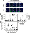
Monitoring oviduct infection following C. muridarum intravaginal inoculation. (A) The oviduct sections from genital tract tissues harvested from mice on days 3 (n = 5 for CBA/J and A/J mice, respectively), 7 (n = 10), 10 (n = 10), 14 (n = 5), and 21 (n = 5) after infection, as described in the legend to Fig. 2, plus additional mice for immunofluorescence assay. The sections were labeled with a DNA dye (blue) and a rabbit antichlamydial antibody (raised with C. muridarum elementary bodies; green). Representative images taken under 10× (a to j) and 100× (a1 to j1) magnification from both CBA/J (a to e and a1 to e1) and A/J (f to j and f1 to j1) at each time as indicated on top of each image are presented with representative inclusions marked with green arrows. White rectangles in images from a 10× objective lens indicate the same areas viewed under the 100× objective lens. (B) The inclusions were counted and semiquantitated as described in Materials and Methods, as shown along the y axis. The number of inclusions per slide varied considerably from 0 to >300 (image a). To show differences between the two groups of mice, the region covering samples with 25 or fewer inclusions was magnified as image a1. Note that on day 7 after infection, significantly more inclusions were detected in the oviduct sections of A/J mice (a1, open diamonds). The number of inclusions rapidly increased in CBA/J mice (solid), surpassing the A/J mice on day 10 postinfection or thereafter. (C) The microscopic observations described here were validated by titrating infectious chlamydial organisms from oviduct/ovary tissue homogenates harvested from mice on days 3, 7, 10, 14, 21, and 28, as indicated along the x axis. The recovered IFUs, expressed in log10 values, are shown along the y axis. Note that the IFUs were higher in A/J oviduct/ovary tissue samples on day 7 but significantly lower on days 14 and 21 (*, P < 0.05; Student t test). The IFU recovery study was carried out with 5 mice per group.

Similar articles

Cited by

References

    1. Sherman KJ, Daling JR, Stergachis A, Weiss NS, Foy HM, Wang SP, Grayston JT. 1990. Sexually transmitted diseases and tubal pregnancy. Sex. Transm. Dis. 17:115–121. 10.1097/00007435-199007000-00001 - DOI - PubMed
    1. de la Maza LM, Pal S, Khamesipour A, Peterson EM. 1994. Intravaginal inoculation of mice with the Chlamydia trachomatis mouse pneumonitis biovar results in infertility. Infect. Immun. 62:2094–2097 - PMC - PubMed
    1. Shah AA, Schripsema JH, Imtiaz MT, Sigar IM, Kasimos J, Matos PG, Inouye S, Ramsey KH. 2005. Histopathologic changes related to fibrotic oviduct occlusion after genital tract infection of mice with Chlamydia muridarum. Sex. Transm. Dis. 32:49–56. 10.1097/01.olq.0000148299.14513.11 - DOI - PubMed
    1. Murthy AK, Li W, Guentzel MN, Zhong G, Arulanandam BP. 2011. Vaccination with the defined chlamydial secreted protein CPAF induces robust protection against female infertility following repeated genital chlamydial challenge. Vaccine 29:2519–2522. 10.1016/j.vaccine.2011.01.074 - DOI - PMC - PubMed
    1. Stephens RS. 2003. The cellular paradigm of chlamydial pathogenesis. Trends Microbiol. 11:44–51. 10.1016/S0966-842X(02)00011-2 - DOI - PubMed

MeSH terms

LinkOut - more resources