

Robotic-assisted surgery approach in a biliary rhabdomyosarcoma misdiagnosed as choledochal cyst
- PMID: 24711907
- PMCID: PMC3977170
- DOI: 10.4081/rt.2014.5173
Robotic-assisted surgery approach in a biliary rhabdomyosarcoma misdiagnosed as choledochal cyst
Abstract
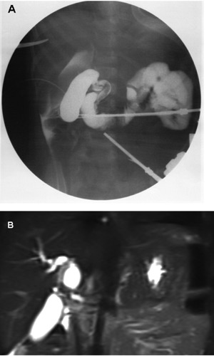
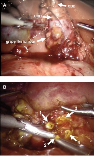
Rhabdomyosarcoma is a soft tissue malignant tumor affecting 1% of children from 0 to 14 years. Preoperative imaging may not always be diagnostic for hepatobiliary rhabdomyosarcoma and differential diagnosis with choledochal cyst (CC) could be difficult. We report a case of 2-years-old girl with a strange CC pattern of presentation. A grapelike lesion involving the choledochal and biliary ducts was easily and completely resected by robotic assisted surgery. Since no previous reports were available about oncologic safety of robotic approach, the porto-enterostomy was performed in open surgery. On histologic examination, the specimen revealed a botryoidembryonal rhabdomyosarcoma affecting both the common bile duct and the common hepatic duct. One year postoperatively the child is safe of tumor relapse. Robotic approach seems to be safe and advantageous to obtain a radical excision of the tumor at the porta hepatis, even in case of misdiagnosed malignant lesion mimicking a CC.
Keywords: biliary botryoid rhabdomyosarcoma; choledochal cyst; porta hepatis; robotic surgery.
Conflict of interest statement
Conflict of interests: the authors declare no potential conflict of interests.
Figures
Similar articles
-
[Rhabdomyosarcoma of the common bile duct mimicking choledochal cyst: a rare cause of obstructive jaundice].Zhongguo Dang Dai Er Ke Za Zhi. 2020 Dec;22(12):1338-1343. doi: 10.7499/j.issn.1008-8830.2007118. Zhongguo Dang Dai Er Ke Za Zhi. 2020. PMID: 33328007 Free PMC article.
-
Laparoscopically assisted extrahepatic bile duct excision with ductoplasty and a widened hepaticojejunostomy for complicated hepatobiliary dilatation.Pediatr Surg Int. 2014 Jun;30(6):593-8. doi: 10.1007/s00383-014-3501-2. Epub 2014 Apr 10. Pediatr Surg Int. 2014. PMID: 24718723
-
Botryoid rhabdomyosarcoma of the biliary tract in children: a unique case report.Eur J Cancer Care (Engl). 2006 Dec;15(5):463-6. doi: 10.1111/j.1365-2354.2006.00683.x. Eur J Cancer Care (Engl). 2006. PMID: 17177904 Review.
-
Hepatobiliary rhabdomyosarcoma mimicking choledochal cyst: Lessons learned.Int J Surg Case Rep. 2014;5(4):196-9. doi: 10.1016/j.ijscr.2014.01.020. Epub 2014 Feb 7. Int J Surg Case Rep. 2014. PMID: 24636980 Free PMC article.
-
Initial experience with complex laparoscopic biliary surgery in children: biliary atresia and choledochal cyst.J Pediatr Surg. 2004 Jun;39(6):804-7; discussion 804-7. doi: 10.1016/j.jpedsurg.2004.02.018. J Pediatr Surg. 2004. PMID: 15185200 Review.
Cited by
-
Laparoscopic management of Rhabdomyosarcoma of common Bile duct, Case report.Ann Med Surg (Lond). 2020 Sep 20;59:118-121. doi: 10.1016/j.amsu.2020.09.023. eCollection 2020 Nov. Ann Med Surg (Lond). 2020. PMID: 33005400 Free PMC article.
-
Pediatric choledochal cysts: diagnosis and current management.Pediatr Surg Int. 2017 Jun;33(6):637-650. doi: 10.1007/s00383-017-4083-6. Epub 2017 Mar 31. Pediatr Surg Int. 2017. PMID: 28364277 Review.
References
-
- Stocker JT. Hepatic tumors in children. Clin Liver Dis 2001;5:259-81 - PubMed
-
- Meyers RL. Tumors of the liver in children. Surg Oncol 2007;16:195-203 - PubMed
-
- Kebudi R, Görgun O, Ayan I, et al. Rhabdmyosarcoma of the biliary tree. Pediatr Int 2003;45:469-71 - PubMed
-
- Tireli GA, Sander S, Dervisoglu S, et al. Embryonal rhabdomyosarcoma of the common bile duct mimicking choledochal cyst. J Hepatobiliary Pancreat Surg 2005;12:263-5 - PubMed
Publication types
LinkOut - more resources
Full Text Sources
Other Literature Sources
