. 2013 Jul 23:2:336.
doi: 10.1186/2193-1801-2-336. eCollection 2013.

Skeletal gene expression in the temporal region of the reptilian embryos: implications for the evolution of reptilian skull morphology

Affiliations

Skeletal gene expression in the temporal region of the reptilian embryos: implications for the evolution of reptilian skull morphology

Masayoshi Tokita et al. Springerplus. .

Abstract

Reptiles have achieved highly diverse morphological and physiological traits that allow them to exploit various ecological niches and resources. Morphology of the temporal region of the reptilian skull is highly diverse and historically it has been treated as an important character for classifying reptiles and has helped us understand the ecology and physiology of each species. However, the developmental mechanism that generates diversity of reptilian skull morphology is poorly understood. We reveal a potential developmental basis that generates morphological diversity in the temporal region of the reptilian skull by performing a comparative analysis of gene expression in the embryos of reptile species with different skull morphology. By investigating genes known to regulate early osteoblast development, we find dorsoventrally broadened unique expression of the early osteoblast marker, Runx2, in the temporal region of the head of turtle embryos that do not form temporal fenestrae. We also observe that Msx2 is also uniquely expressed in the mesenchymal cells distributed at the temporal region of the head of turtle embryos. Furthermore, through comparison of gene expression pattern in the embryos of turtle, crocodile, and snake species, we find a possible correlation between the spatial patterns of Runx2 and Msx2 expression in cranial mesenchymal cells and skull morphology of each reptilian lineage. Regulatory modifications of Runx2 and Msx2 expression in osteogenic mesenchymal precursor cells are likely involved in generating morphological diversity in the temporal region of the reptilian skull.

Keywords: Development; Heterotopy; Morphology; Osteogenesis; Reptiles; Skull.


Figures
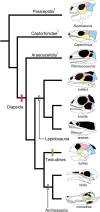
Figure 1
Figure 1
Simplified phylogeny of the Reptilia highlighting diversity of their skull morphology. Paleontological evidence suggests that all reptiles, including extant lizards, snakes, tuatara, crocodiles, birds, and turtles, were derived from ancestor whose temporal region was completely roofed by bone. Earliest diapsid reptiles such as Petrolacosaurus (Araeoscelidia) acquired two temporal openings (fenestrae) on either side of the skull (red vertical bar). Recent molecular phylogenies indicate that turtles (Testudines) were derived from diapsid ancestor, which would require secondary closure of temporal fenestrae (green vertical bar). The bone surrounding anteroventral border of the upper temporal fenestra and anterodorsal border of the lower temporal fenestra in diapsids: the postorbital was colored in pink. The bone surrounding posteroventral border of the upper temporal fenestra and posterodorsal border of the lower temporal fenestra in diapsids: the squamosal was colored in blue. The bone surrounding the anteroventral margin of the lower temporal fenestra in diapsids: the jugal was colored in yellow. Lizards do not have the lower temporal bar. Both upper and lower temporal bars are absent in snakes and birds. Other extinct diapsid lineages such as Ichthyosauria and Sauropterygia were not included in phylogeny for simplicity.
Figure 2
Figure 2
Expression of musculoskeletal tissue marker genes in the head of crocodile embryos at stage 15. (A) Lateral view of the head of a crocodile embryo at stage 15. (B-D) Frontal sections prepared around the plane indicated by the red line in (A). (E-G) Frontal sections prepared around the plane indicated by the blue line in (A). (H) Frontal sections prepared around the plane indicated by the green line in (A). (B, E, and H) Runx2-positive mesenchymal cells are observed at the location where future dermatocranial elements are developed. (C and F) Expression of MyoD is detected at the cranial muscular tissues. (D and G) Cartilaginous tissues, including the braincase, quadrate, and Meckel's, are clearly labeled by Sox9 probe. The red outlined domains in (B, D, E, and G) indicate the location of the pseudotemporal muscle (ptm) deduced from adjacent sections where muscular tissues are labeled by MyoD probe. Scale bar in (A) is 1 mm. Scale bars in (B-H) are 0.5 mm.
Figure 3
Figure 3
Expression of musculoskeletal tissue marker genes in the head of crocodile embryos at stage 17. (A) Lateral view of the head of a crocodile embryo at stage 17. (B-D) Frontal sections prepared around the plane indicated by the red line in (A). (E-I) Frontal sections prepared around the plane indicated by the blue line in (A). (B and E) Expression of Runx2 is more concentrated to the precursors of dermatocranial elements, compared to the previous stages. (C, D, F, and G) Cranial muscular and cartilaginous tissues are clearly labeled by MyoD and Sox9 probes, respectively. (H and I) Expression domains of Scx and Six2 are indicated by arrowheads. The former is expressed in tendinous tissues accompanying cranial muscles and the latter is expressed mainly in connective tissue cells associated with cartilages of the jaw and the braincase. The red outlined domains in (B, D, E, G, H, and I) indicate the location of the pseudotemporal muscle deduced from adjacent sections where muscular tissues are labeled by MyoD probe. Green line in (A) indicates the plane where sections given in Figure 7D, E and F were prepared. Scale bar in (A) is 1 mm. Scale bars in (B-I) are 0.5 mm.
Figure 4
Figure 4
Expression of musculoskeletal tissue marker genes in the head of turtle embryos at stage 15. (A) Lateral view of the head of a turtle embryo at stage 15. (B-F) Frontal sections prepared around the plane indicated by the red line in (A). (G-I) Frontal sections prepared around the plane indicated by the blue line in (A). (B, G) Runx2-positive mesenchymal cells (arrows) are broadly distributed at lateral portion of the head, from the top of the head to the ventral margin of the first pharyngeal arch. (C and H) Cranial muscular tissues are clearly labeled by MyoD probe. (D and I) Cartilaginous tissues, including the braincase and quadrate, are clearly labeled by Sox9 probe. (E) Scx is expressed in tendon primordia accompanying cranial muscles (arrowheads). (F) Six2 is expressed mainly in connective tissue cells associated with the braincase cartilage and jaw adductor muscle (arrowheads). Note that the anlage of jaw adductor muscle (red outlined domain) is covered by a thick layer of Runx2-positive mesenchyme laterally. Scale bar in (A) is 1 mm. Scale bars in (B-I) are 0.5 mm.
Figure 5
Figure 5
Expression of musculoskeletal tissue marker genes in the head of turtle embryos at stage 17. (A) Lateral view of the head of a turtle embryo at stage 17. (B-F) Frontal sections prepared around the plane indicated by the blue line in (A). (B) Runx2-positive mesenchymal cells (arrows) are broadly distributed at the lateral portion of the head, from the top of the head to the ventral margin of the first pharyngeal arch derivative. (C) Cranial muscular tissues are clearly labeled by MyoD probe. (D) Cartilaginous tissues, including the braincase and quadrate, are clearly labeled by Sox9 probe. (E) Scx is expressed in tendinous tissues at the periphery of external adductor muscle (ame) (arrowheads) and in the bodenaponeurosis (boap. central tendon of jaw adductor muscle). (F) Six2 is expressed mainly in connective tissue cells associated with cartilages of the braincase and quadrate, as well as in connective tissue cells within jaw muscles (arrowheads). Note that the external adductor muscle (red outlined domain) is covered by a thick layer of Runx2-positive mesenchyme laterally. Scale bar in (A) is 1 mm. Scale bars in (B-F) are 0.5 mm.
Figure 6
Figure 6
Expression of Bmp4 , Msx1 , and Msx2 in crocodile and turtle embryos at stage 15. (A-D) Frontal sections prepared around the plane indicated by the red line in Figure 2A. (E, F) Frontal sections prepared around the plane indicated by the blue line in Figure 2A. (G-J) Frontal sections prepared around the plane indicated by the red line in Figure 4A. (K, L) Frontal sections prepared around the plane indicated by the blue line in Figure 4A. (A and G) In both crocodile and turtle embryos, expression of Bmp4 is detected at the mesenchyme distributed in medial part of the jaw primordia (arrows). (B and H) Expression of Msx1 is detected at the mesenchyme that occupies the domain close to jaw articulation and lateral to quadrate and Meckel's cartilages (arrows). (C and E) In crocodile embryos, Msx2 is expressed in a thin layer of mesenchymal cells surrounding dorsal aspect of the brain and in a population of the mesenchyme that occupies the domain between ventrolateral part of quadrate cartilage and surface epidermis (arrows). (I and K) In turtle embryos, Msx2 is expressed in mesenchymal cells that populate lateral aspect of the head (arrows). In contrast to the condition in crocodile embryos, the ventral edge of Msx2-expressing mesenchymal layer is terminated ventral to the eye in turtle embryos and these cells cover MyoD-expressing jaw adductor muscle precursor (J and L) laterally. The red outlined domains in (A-C, and E) indicate the location of the anlagen of the pseudotemporal muscle deduced from adjacent sections where muscular tissues are labeled by MyoD probe. The red outlined domains in (G-I, and K) indicate the location of the anlagen of jaw adductor muscle. Scale bars are 0.5 mm.
Figure 7
Figure 7
Expression of Bmp4 , Msx1 , and Msx2 in crocodile and turtle embryos at stage 17. (A-C) Frontal sections prepared around the plane indicated by the blue line in Figure 3A. (D-F) Sections prepared around the plane indicated as the green line in Figure 3A. (G-I) Sections prepared around the plane indicated as the red line in Figure 5A. (J) Section prepared around the plane indicated as the blue line in Figure 5A. (A, D, and G) In both crocodile and turtle embryos, expression of Bmp4 is detected in the epithelium of the cochlear canal, the mesenchyme surrounding the eye, and the mesenchyme distributed in medial part of the jaw (arrows). (B, E, and H) Expression of Msx1 is detected at the mesenchymal cells that later differentiates into quadratojugal bone (qj) and in a thin layer of mesenchymal cells that covers brain dorsally (arrows at the top of E and H), as well as in the epithelium of the cochlear canal. (C and F) In crocodile embryos, expression of Msx2 is detected at a population of mesenchymal cells in close proximity of postorbital and quadratojugal bones, as well as in a layer of the mesenchyme surrounding the brain dorsally where future a pair of parietal bones are developed. (I and J) In turtle embryos, Msx2 was expressed in a thick layer of mesenchymal cells that populate lateral aspect of the head (arrows). The Msx2-positive mesenchymal cells cover the external adductor muscle precursors (red outlined domains in G- J) laterally. The red outlined domains in (A-F) indicate the location of the pseudotemporal muscle deduced from adjacent sections where muscular tissues are labeled by MyoD probe. The blue outlined domains in (A-J) indicate the location of quadrate cartilage deduced from adjacent sections where cartilaginous tissues are labeled by Sox9 probe. Scale bars are 0.5 mm.
Figure 8
Figure 8
Expression of the genes that regulate the development of cranial musculoskeletal tissues in snake embryos. (A) Lateral view of the head of a snake embryo at stage 26. (B-E) Frontal sections prepared around the plane indicated by the red line in (A). (F-I) Frontal sections around the blue line in (A). (J) The head of a snake embryo at stage 29. (K-N) Frontal sections around the red line in (J). (O-R) Frontal sections around the blue line in (J). (S) The head of a snake embryo at stage 31. (T-W) Frontal sections around the red line in (S). (B, F, K, O, and T) Expression of Runx2. (C, G, L, P, and U) Expression of MyoD. (D, H, M, Q, and V) Expression of Sox9. (E, I, N, R, and W) Expression of Msx2. In snake embryos, expression of Runx2 and Msx2 are detected in the precursor cells of dermatocranial elements (arrows), as in crocodile and turtle embryos. However, these mesenchymal cells are not seen in the temporal region lateral to the jaw adductor muscles. Instead, these cells are distributed in the vicinity of the brain, laterally covering it, and differentiate into the parietal bone in older embryos. Open arrowheads in (F, K, N, O, R, T, and W) indicate the mesenchyme around Sox9-positive jaw cartilages, where the expression of Runx2 and Msx2 is detected. The red outlined domains in (O, Q, R, T, V, and W) indicate the location of the external adductor muscle deduced from adjacent sections where muscular tissues are labeled by MyoD probe. Scale bars in (A), (J), and (S) are 1 mm. Scale bars in other pictures are 0.5 mm.
Figure 9
Figure 9
Potential developmental basis that generates morphological diversity in the temporal region of the reptilian skull. All extant reptilian lineages are considered to be derived from ancestor with diapsid skull. In crocodiles that have both upper and lower temporal bars like the stem Diapsida (e.g., Petrolacosaurus), osteogenic mesenchymal precursor cells which express Runx2 and/or Msx2 are distributed at the domain where future temporal bars are formed in the head of early stage embryo (top of the middle column). Through ontogeny (black arrow), these osteoblast precursors may differentiate into the dermatocranial elements including upper and lower temporal bars (bottom of the middle column). Between these bony bars, both upper and lower temporal fenestrae are clearly recognized. In turtles (left white arrow), distribution of osteogenic mesenchymal precursor cells is broadened in a dorsal-ventral direction, filling the whole lateral portion of the head of early embryos (top of the left column). Through ontogeny, these osteoblast precursors may differentiate into the dermatocranial elements roofing the temporal region of the head (bottom of the left column). In snakes that have modified diapsid skull where temporal bars are absent, osteogenic mesenchymal precursor cells do not fill lateral domain of the head of embryo. Rather, these cells are mainly distributed in the vicinity of the brain (top of the right column). Through ontogeny, these osteoblast precursors may differentiate into the dermatocranial elements accompanying the braincase, without forming bony temporal bars (bottom of the right column). A condensed mesenchymal layer that differentiates into the braincase in later stages is highlighted by dotted line in the head of embryos.

Similar articles

Cited by

References

    1. Abzhanov A, Rodda SJ, McMahon AP, Tabin CJ. Regulation of skeletogenic differentiation in cranial dermal bone. Development. 2007;134:3133–3144. doi: 10.1242/dev.002709. - DOI - PubMed
    1. Antonopoulou I, Mavrogiannis LA, Wilkie AOM, Morriss-Kay GM. Alx4 and Msx2 play phenotypically similar and additive roles in skull vault differentiation. J Anat. 2004;204:487–499. doi: 10.1111/j.0021-8782.2004.00304.x. - DOI - PMC - PubMed
    1. Bell DM, Leung KKH, Wheatley SC, Ng LJ, Zhou S, Ling KW, Sham MH, Koopman P, Tam PPL, Cheah KSE. Sox9 directly regulates the type-II collagen gene. Nat Genet. 1997;16:174–178. doi: 10.1038/ng0697-174. - DOI - PubMed
    1. Benton MJ. Vertebrate palaeontology 3rd edition. Malden: Blackwell Publishing; 2005.
    1. Bobola N, Carapuco M, Ohnemus S, Kanzler B, Leibbrandt A, Neubuser A, Drouin J, Mallo M. Mesenchymal patterning by Hoxa2 requires blocking Fgf-dependent activation of Ptx1. Development. 2003;130:3403–3414. doi: 10.1242/dev.00554. - DOI - PubMed

LinkOut - more resources