

Human internal jugular valve M-mode ultrasound characterization
- PMID: 24712644
- PMCID: PMC4031920
- DOI: 10.2174/1567202611666140408094014
Human internal jugular valve M-mode ultrasound characterization
Abstract
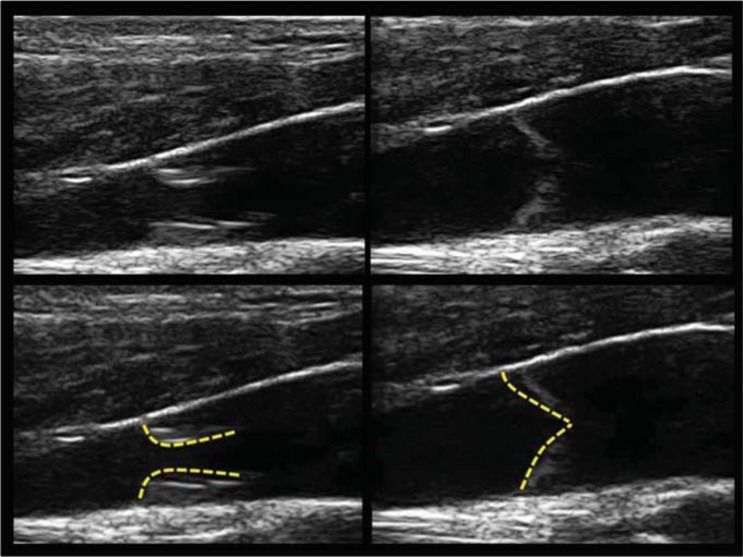
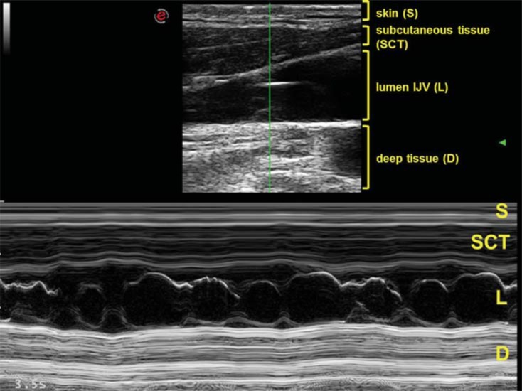
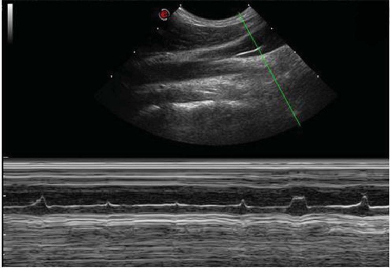
In humans the mechanism governing the internal jugular vein (IJV) valve opening and closure is still unclear. M-mode is used in echo-cardiology for the heart valves assessment. Sometimes it was performed also in deep peripheral veins and in vena cava assessment, but never in the IJV valve. Aim of the present study is to investigate the IJV valves physiology in healthy volunteers, by means of both B and M-mode ultrasound. Eighty-three (83) healthy volunteers (35 Male, 48 Female, 25.7±6.7 y.o.), for a total of 166 IJVs, were enrolled. The entire cohort underwent IJVs high-resolution B and M-mode evaluation, in standardized postural and respiratory conditions. Presence, motility, and number of cusps, as well as their opening and closure mechanism have been assessed. Bilateral valve absence occurred in 13/83 (16%), whereas at least a one side absence was recorded in 38/83 (46% of the cohort) (p<0.0356). Valve leaflets were always mobile and respectively bi-cusps in 34%, or mono-cusp in 27%. The latter was significantly more frequent on the left side (35%) than on the right side (19%) (p<0.0013). In supine, M-mode valve opening was synchronous with the cardiac cycle. To the contrary, in an upright position, the valve remained always open and saddled to the wall, independently from the cardiac cycle. In healthy subjects, the IJV valve leaflets are always mobile, but the significant rate of mono and bilateral absence could suggest a progressive phylogenetic importance loss of such apparatus. M-mode ultrasound enhances the characterization of IJV valve, for this reason it should be taken into consideration to routinely add it to the cerebral venous return investigation.
Figures

References
-
- Fisher J. Jugular venous valves and,physical signs. Chest. 1984;85(5):85–6. - PubMed
-
- Brownlow RL Jr, McKinney WM. Ultrasonic evaluation of jugular venous valve competence. J Ultrasound Med. 1985;4(4):169–72. - PubMed
-
- Lepori D, Capasso P, Fournier D, Genton CY, Schnyder P. Hight-resolution ultrasound evaluation of internal jugular venous valves. EurRadiol. 1999;9(6):1222–6. - PubMed
-
- Valecchi D, Bacci D, Gulisano M , et al. Internal jugular vein valves: an assessment of prevalence. morphology and competence by color Doppler echography in 240 healthy subjects. IJAE. 2010;115(3):185–9. - PubMed
-
- Zamboni P, Menegatti E, Pomidori L , et al. Does thoracic pump influence the cerebral venous return?. J Appl Physiol. 2012;112(5):904–10. - PubMed
Publication types
MeSH terms
LinkOut - more resources
Full Text Sources
Other Literature Sources