. 2014 Apr;39(7):1159-69.
doi: 10.1111/ejn.12422.

BDNF contributes to both rapid and homeostatic alterations in AMPA receptor surface expression in nucleus accumbens medium spiny neurons

Affiliations

BDNF contributes to both rapid and homeostatic alterations in AMPA receptor surface expression in nucleus accumbens medium spiny neurons

Jeremy M Reimers et al. Eur J Neurosci. 2014 Apr.

Abstract

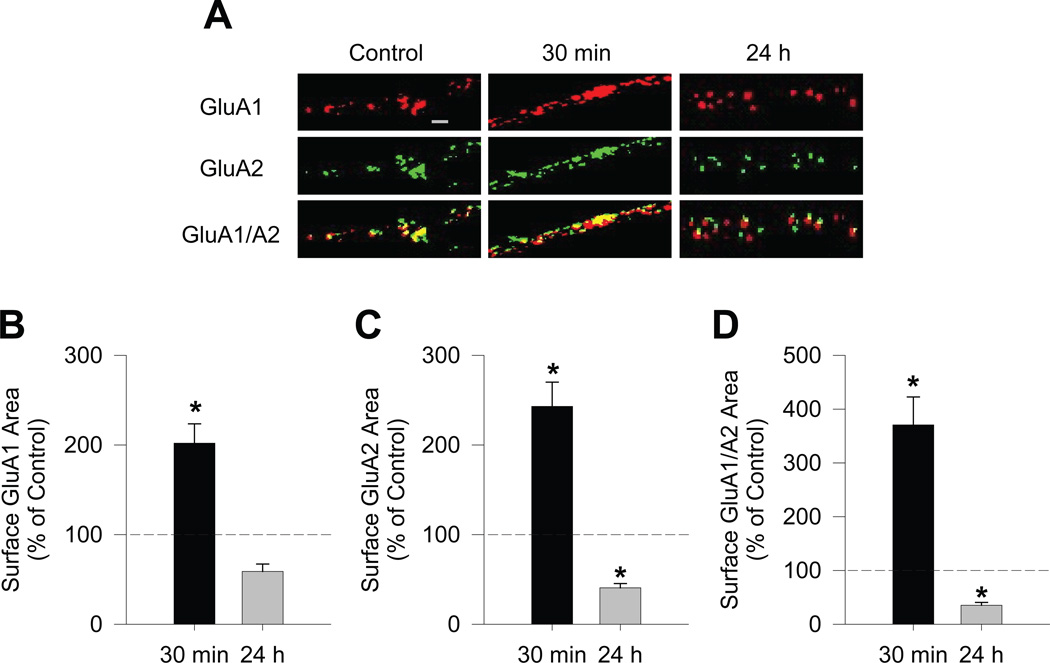
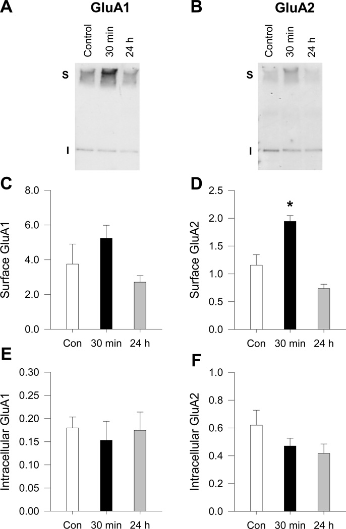
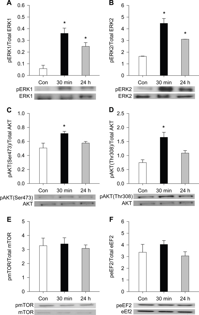
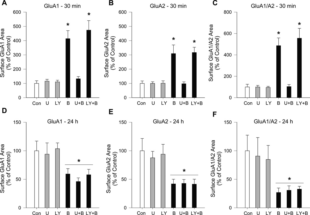
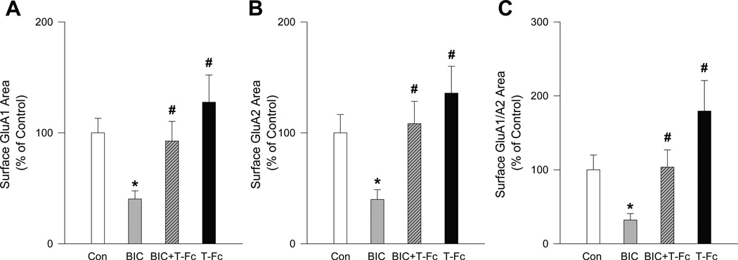
Brain-derived neurotrophic factor (BDNF) plays a critical role in plasticity at glutamate synapses and in the effects of repeated cocaine exposure. We recently showed that intracranial injection of BDNF into the rat nucleus accumbens (NAc), a key region for cocaine addiction, rapidly increases α-amino-3-hyroxy-5-methyl-4-isoxazole-propionic acid receptor (AMPAR) surface expression. To further characterize BDNF's role in both rapid AMPAR trafficking and slower, homeostatic changes in AMPAR surface expression, we investigated the effects of acute (30 min) and long-term (24 h) treatment with BDNF on AMPAR distribution in NAc medium spiny neurons from postnatal rats co-cultured with mouse prefrontal cortex neurons to restore excitatory inputs. Immunocytochemical studies showed that acute BDNF treatment increased cell surface GluA1 and GluA2 levels, as well as their co-localization, on NAc neurons. This effect of BDNF, confirmed using a protein crosslinking assay, was dependent on ERK but not AKT signaling. In contrast, long-term BDNF treatment decreased AMPAR surface expression on NAc neurons. Based on this latter result, we tested the hypothesis that BDNF plays a role in AMPAR 'scaling down' in response to a prolonged increase in neuronal activity produced by bicuculline (24 h). Supporting this hypothesis, decreasing BDNF signaling with the extracellular BDNF scavenger TrkB-Fc prevented the scaling down of GluA1 and GluA2 surface levels in NAc neurons normally produced by bicuculline. In conclusion, BDNF exerts bidirectional effects on NAc AMPAR surface expression, depending on duration of exposure. Furthermore, BDNF's involvement in synaptic scaling in the NAc differs from its previously described role in the visual cortex.

Keywords: co-culture; mouse; rat; receptor trafficking; synaptic scaling.


Conflict of interest statement

The authors declare no conflict of interest.

Figures

Figure 1
Figure 1
Surface expression and co-localization of GluA1 and GluA2 on NAc medium spiny neurons are robustly increased after acute BDNF treatment and decreased after long-term BDNF treatment of NAc/PFC co-cultures. (A) Representative images of GluA1, GluA2 and colocalized GluA1/A2 immunostaining on medium spiny neurons in NAc/PFC co-cultures. Co-cultures were treated with BDNF (25ng/mL) for 30 min or 24 h. Surface GluA1 (red) and GluA2 (green) were detected using live cell labeling. Scale bar, 2.5μm. (B-D) Quantification of the area of cell surface GluA1 (B), GluA2 (C), and GluA1/A2 co-localization (D) (n = 18–26 cells/group). Results are shown as area of surface staining (mean + SEM), normalized to the mean of the vehicle control group, which is indicated by the dashed line (control group: GluA1, 100 ± 14.67%; GluA2, 100 ± 10.23%; GluA1/A2, 100 ± 18.12%). Data were analyzed using a one-way ANOVA on ranks followed by a Dunn’s test if group differences were found. *P < 0.05 vs. control.
Figure 2
Figure 2
BS3 crosslinking followed by Western blotting reveals that acute BDNF treatment increases AMPAR surface expression in high-density NAc cultures. NAc cultures were treated with BDNF (25ng/mL) for 30 min or 24 h. Following BDNF treatment, surface proteins were crosslinked using BS3 and Western blotting was used to quantify surface (S) and intracellular (I) levels of AMPAR subunits. (A–B) Representative blots of GluA1 (left) and GluA2 (right) show the migration pattern for the AMPAR subunits. BS3 crosslinked surface proteins migrate at a higher molecular weight than the intracellular pool, which represents unmodified receptors (~100kDa). (C-F) Effect of 30 min or 24 h incubation with BDNF on surface GluA1 (C), surface GluA2 (D), intracellular GluA1 (E), and intracellular GluA2 (F) (n = 4 wells/group). Error bars represent SEM. Data were analyzed using a one-way ANOVA followed by a Dunnett’s test if group differences were found. *P < 0.05 vs. control.
Figure 3
Figure 3
Effect of BDNF treatment on phosphorylation of ERK1, ERK2, AKT, mTOR and eEF2 in high-density NAc cultures. These proteins were selected for analysis because they are downstream of BDNF-TrkB signaling. NAc cultures were stimulated with BDNF (25ng/mL) for 30 min or 24 h. Western blotting was used to determine the level of protein phosphorylation. The 30 min BDNF treatment increased phosphorylation of ERK1 (A), ERK2 (B), AKT (Ser 473) (C), and AKT (Thr 308) (D), but not mTOR (E), and eEF2 (F). The 24 h BDNF treatment only increased ERK1 and ERK2 phosphorylation. All data are shown as phospho-protein normalized to the total level of target protein determined with a phosphorylation-independent antibody. Representative blots are shown below the graphs. Error bars represent SEM (n = 4 wells/group). Data were analyzed using a one-way ANOVA followed by a Dunnett’s test if group differences were found. *P < 0.05 vs. control.
Figure 4
Figure 4
The increase in surface AMPAR levels induced by acute BDNF treatment of NAc/PFC co-cultures requires MAPK activation but not the PI3 kinase pathway, whereas effects of long-term BDNF are not altered by inhibitors of either pathway. Inhibition of the MAPK pathway with U0126 (U) prevented the increase in GluA1 and GluA2 surface expression and co-localization observed after 30 min of BDNF (B) treatment, while inhibition of the PI3 kinase pathway with LY294002 (LY) had no effect (panels A-C). In contrast, the percent decrease in AMPAR subunit surface expression produced by 24 h of BDNF treatment did not differ between BDNF, U+BDNF or LY+BDNF groups (D-F). For these experiments, NAc/PFC co-cultures were treated with vehicle, BDNF (25ng/mL), U (10μM), LY (25μM), U+BDNF, or LY+BDNF for 30 min (A-C) or 24 h (D–F) (n = 17–25 cells/group). Surface GluA1 and GluA2 were measured using live cell staining. Results are presented as the area (mean + SEM) of surface GluA1 (A,D), GluA2 (B,E) or GluA1/A2 co-localization (C,F) for each time-point. Data were analyzed using a one-way ANOVA on ranks followed by a Dunn’s test. Analysis for 30 min experiment: *P < 0.05, B and LY+B groups vs. Control. Analysis for 24 h experiment: *P < 0.05, all BDNF groups (B, U+B, LY+B) vs. all non-BDNF exposed groups (Con, U, LY).
Figure 5
Figure 5
BDNF mediates synaptic “scaling down” of AMPARs produced by a prolonged increase in excitatory transmission. Long-term treatment (24 h) of NAc/PFC co-cultures with the GABAA receptor bicuculline (BIC; 20 µM) produced a significant decrease in surface expression of GluA1 (A) and GluA2 (B) as well as GluA1/A2 co-localization (C). Co-incubation with the membrane-impermeable extracellular scavenger of BDNF TrkB-Fc (T-Fc, 0.5 µg/mL) prevented the decrease in GluA1 (A), GluA2 (B) and GluA1/A2 co-localization (C) normally produced by BIC, while 24 h treatment with T-Fc alone produced trends towards increased AMPAR surface expression. Results are shown as area of surface staining (mean + SEM), normalized to the mean of the respective vehicle control group. Data were analyzed using a one-way ANOVA on ranks followed by a Dunn’s test if group differences were found. * P < 0.05 vs. control, # P < 0.05 vs. BIC.

Similar articles

Cited by

References

    1. Almeida RD, Manadas BJ, Melo CV, Gomes JR, Mendes CS, Grãos MM, Carvalho RF, Carvalho AP, Duarte CB. Neuroprotection by BDNF against glutamate-induced apoptotic cell death is mediated by ERK and PI3-kinase pathways. Cell Death Differ. 2005;12:1329–1343. - PubMed
    1. Altar CA, Cai N, Bliven T, Juhasz M, Conner JM, Acheson AL, Lindsey RM, Wiegland SJ. Anterograde transport of brain-derived neurotrophic factor and its role in the brain. Nature. 1997;389:856–860. - PubMed
    1. Anggono V, Huganir RL. Regulation of AMPA receptor trafficking and synaptic plasticity. Curr. Opin. Neurobiol. 2012;22:461–469. - PMC - PubMed
    1. Bahi A, Boyer F, Dreyer J-L. Role of accumbens BDNF and TrkB in cocaine-induced psychomotor sensitization, conditioned place preference and reinstatement in rats. Psychopharmacology. 2008;199:169–182. - PubMed
    1. Berglind WJ, See RE, Fuchs RA, Ghee SM, Whitfield TW, Jr, Miller SW, McGinty JF. A BDNF infusion into the medial prefrontal cortex suppresses cocaine seeking in rats. Eur. J. Neurosci. 2007;26:757–766. - PubMed

Publication types