

Condylar response to functional therapy with Twin-Block as shown by cone-beam computed tomography
- PMID: 24713070
- PMCID: PMC8638489
- DOI: 10.2319/112713-869.1
Condylar response to functional therapy with Twin-Block as shown by cone-beam computed tomography
Abstract
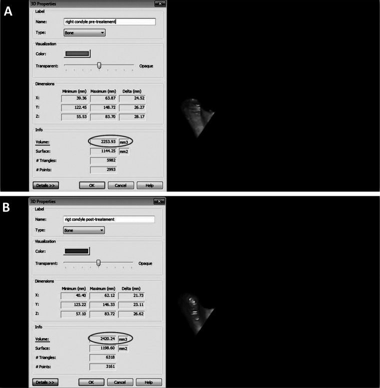
Objective: To evaluate the condylar changes through cone-beam computed tomography (CBCT) images in patients treated with Twin-Block functional appliance.
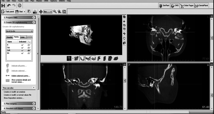
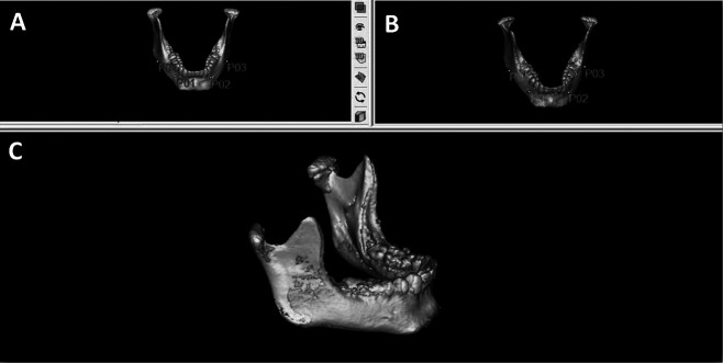
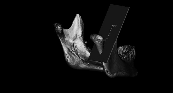
Materials and methods: In this retrospective study, CBCT images of 30 patients who were treated with the Twin-Block appliance were used. Mandible was segmented and pretreatment and posttreatment (T0 and T1) condylar volume was compared. The angle between sella-nasion-Point A (SNA), angle between sella-nasion-Point B (SNB), angle between Point A-nasion-Point B (ANB), midfacial length (Co-A), mandibular length (Co-Gn), and the distances from right condylion to left condylion (CoR-CoL) were also measured on three-dimensional images. Differences were analyzed with Wilcoxon signed rank tests, and Mann-Whitney U-tests were used to compare the scores of male and female participants. Significance was set at P < .05.
Results: In this study, a decrease in SNA and ANB (P < .05 and P < .01, respectively) and an increase in SNB (P < .01) were found. Additionally, CoR-CoL, Co-Gn, and condylar volume increased at both the left and right sides (P < .01). However, increase at Co-A was not statistically significant (P > .05). Comparison of differences by sex was not statistically significant for all measurements (P > .05).
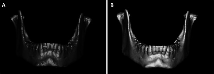
Conclusion: Twin-Block appliance increases condylar volume, mandibular length, and intercondylar distance by stimulating growth of condyle in an upward and backward direction.
Keywords: Condyle; Cone-beam computed tomography; Functional orthopedic appliance; Temporomandibular joint; Twin-Block.
Figures
Similar articles
-
Upper airway changes after Xbow appliance therapy evaluated with cone beam computed tomography.Angle Orthod. 2014 Jul;84(4):693-700. doi: 10.2319/072213-533.1. Epub 2013 Dec 16. Angle Orthod. 2014. PMID: 24328912 Free PMC article.
-
Spatial analysis of condyle position according to sagittal skeletal relationship, assessed by cone beam computed tomography.Prog Orthod. 2013 Oct 18;14:36. doi: 10.1186/2196-1042-14-36. Prog Orthod. 2013. PMID: 24325842 Free PMC article.
-
Three-dimensional skeletal, dentoalveolar and temporomandibular joint changes produced by Twin Block functional appliance.J Orofac Orthop. 2018 Jul;79(4):245-258. doi: 10.1007/s00056-018-0137-1. Epub 2018 Apr 16. J Orofac Orthop. 2018. PMID: 29663034 English.
-
Comparison of Twin Block appliance and Herbst appliance in the treatment of Class II malocclusion among children: a meta-analysis.BMC Oral Health. 2024 Feb 26;24(1):278. doi: 10.1186/s12903-024-04027-w. BMC Oral Health. 2024. PMID: 38409017 Free PMC article.
-
Analysis of efficacy of functional appliances on mandibular growth.Am J Orthod Dentofacial Orthop. 2002 Nov;122(5):470-6. doi: 10.1067/mod.2002.126730. Am J Orthod Dentofacial Orthop. 2002. PMID: 12439474 Review.
Cited by
-
Genetics of the dentofacial variation in human malocclusion.Orthod Craniofac Res. 2015 Apr;18 Suppl 1(0 1):91-9. doi: 10.1111/ocr.12083. Orthod Craniofac Res. 2015. PMID: 25865537 Free PMC article.
-
Comparison of the effects of rapid maxillary expansion versus Twin Block appliance on mandibular growth in skeletal Class II patients.BMC Oral Health. 2020 Dec 1;20(1):350. doi: 10.1186/s12903-020-01344-8. BMC Oral Health. 2020. PMID: 33261594 Free PMC article.
-
3D Comparison of Mandibular Response to Functional Appliances: Balters Bionator versus Sander Bite Jumping.Biomed Res Int. 2018 Apr 24;2018:2568235. doi: 10.1155/2018/2568235. eCollection 2018. Biomed Res Int. 2018. PMID: 29854734 Free PMC article.
-
Assessment of upper airway and temporomandibular joint changes in growing patients with Class II Division 1 malocclusion, treated with the Twin Block appliance: a retrospective cone-beam computed tomography study.Arch Craniofac Surg. 2025 Jun;26(3):92-101. doi: 10.7181/acfs.2024.0093. Epub 2025 Jun 20. Arch Craniofac Surg. 2025. PMID: 40624972 Free PMC article.
-
Three-dimensional spatial analysis of temporomandibular joint in adolescent Class II division 1 malocclusion patients: comparison of Twin-Block and clear functional aligner.Head Face Med. 2024 Jan 6;20(1):4. doi: 10.1186/s13005-023-00404-y. Head Face Med. 2024. PMID: 38184631 Free PMC article.
References
-
- Pancherz H, Ruf S, Kohlhas P. Effective condylar growth and chin position changes in Herbst treatment: a cephalometric roentgenographic long-term study. Am J Orthod Dentofacial Orthop. 1998;114:437–446. - PubMed
-
- Ruf S, Baltromejus S, Pancherz H. Effective condylar growth and chin position changes in activator treatment: a cephalometric roentgenographic study. Angle Orthod. 2001;71:4–11. - PubMed
-
- Pancherz H, Fischer S. Amount and direction of temporomandibular joint growth changes in Herbst treatment: a cephalometric long-term investigation. Angle Orthod. 2003;73:493–501. - PubMed
-
- Dixon D. Radiographic diagnosis of temporomandibular disorders. In: Sadowsky PL, Laskin DM, editors. Seminars in Orthodontics Temporomandibular Joint Disorders Fact or Fiction. Philadelphia, Pa: WB Saunders; 1995. pp. 207–221. - PubMed
-
- Uematsu H, Ichida T, Masumi S, et al. Diagnostic image analyses of activator treated temporomandibular joint in growth and maturing stages. Cranio. 2002;20:254–263. - PubMed
Publication types
MeSH terms
LinkOut - more resources
Full Text Sources
Other Literature Sources
Research Materials
