. 2014 May;124(5):2099-112.
doi: 10.1172/JCI71643. Epub 2014 Apr 8.

Vascular rarefaction mediates whitening of brown fat in obesity

Vascular rarefaction mediates whitening of brown fat in obesity

Ippei Shimizu et al. J Clin Invest. 2014 May.

Abstract

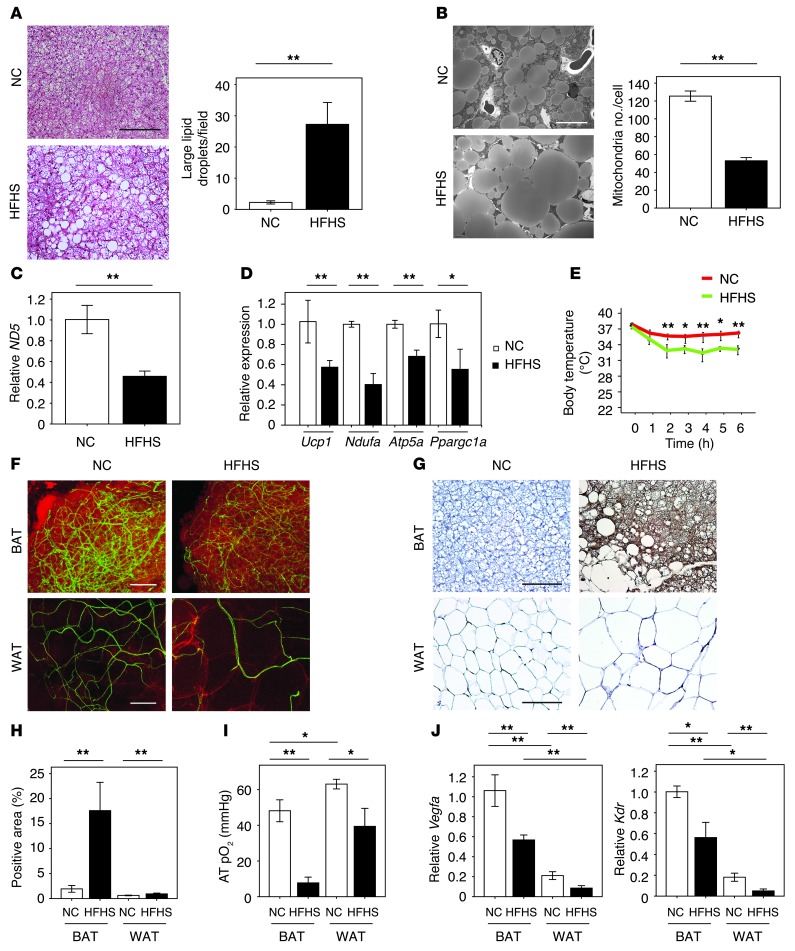
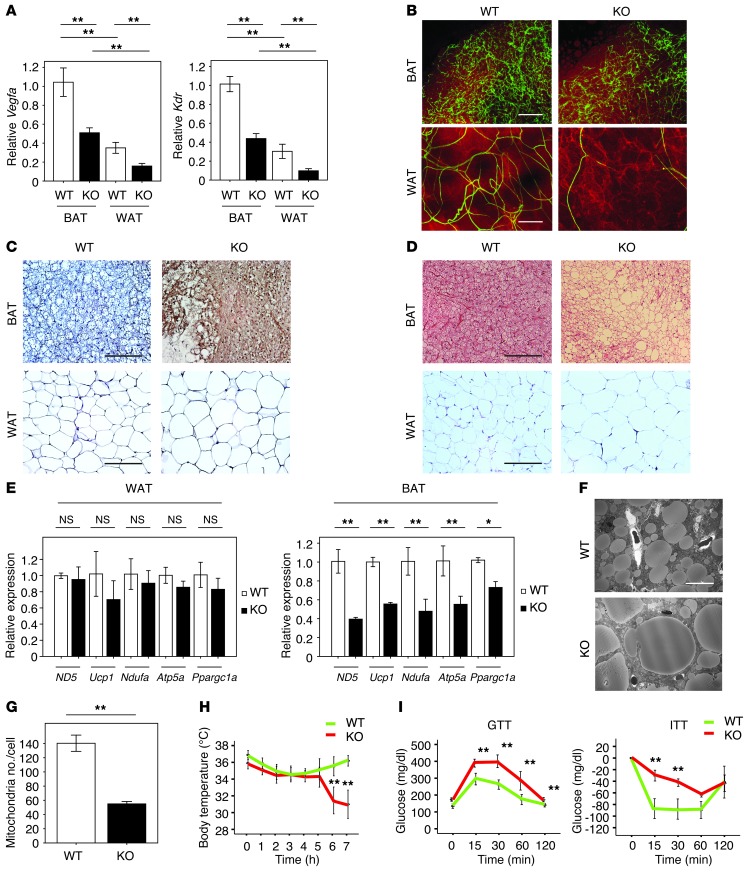
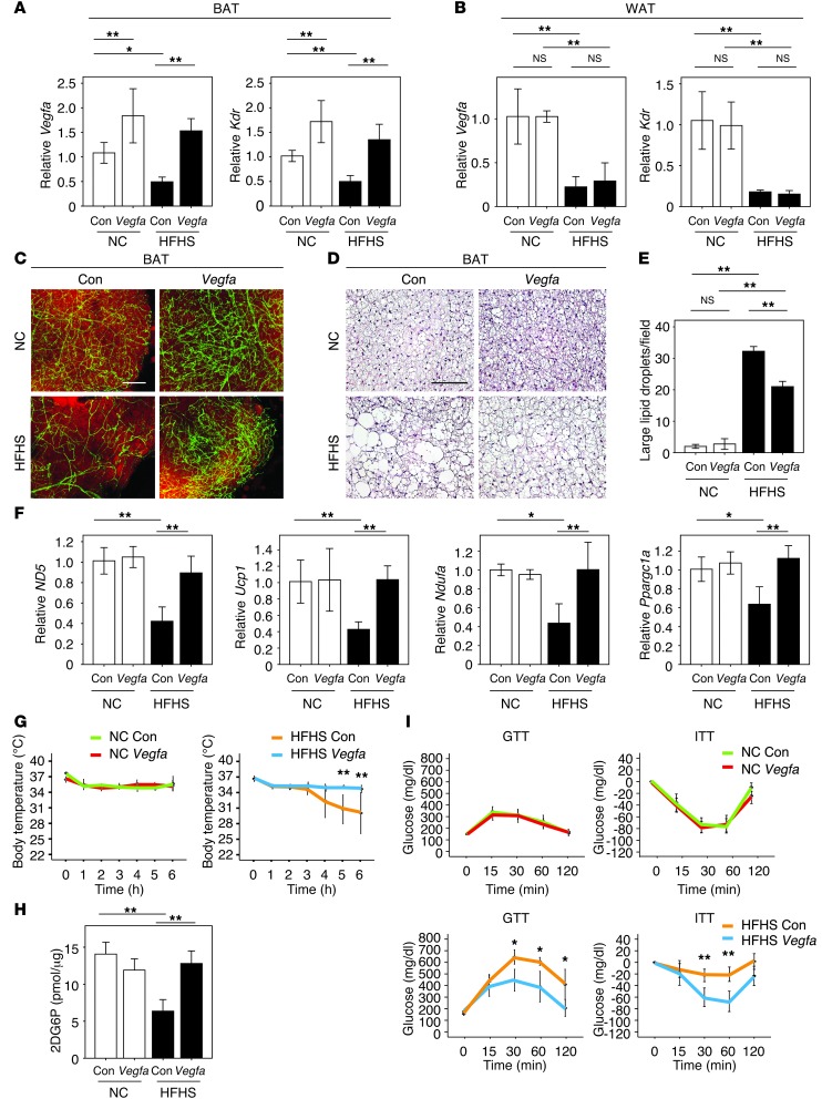
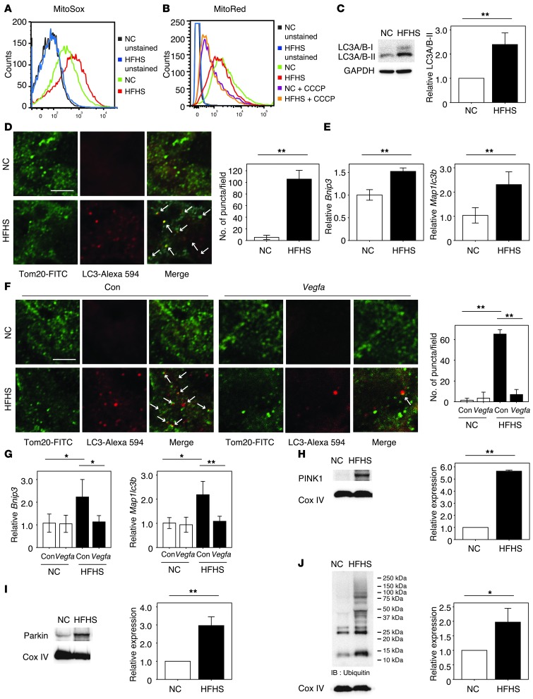
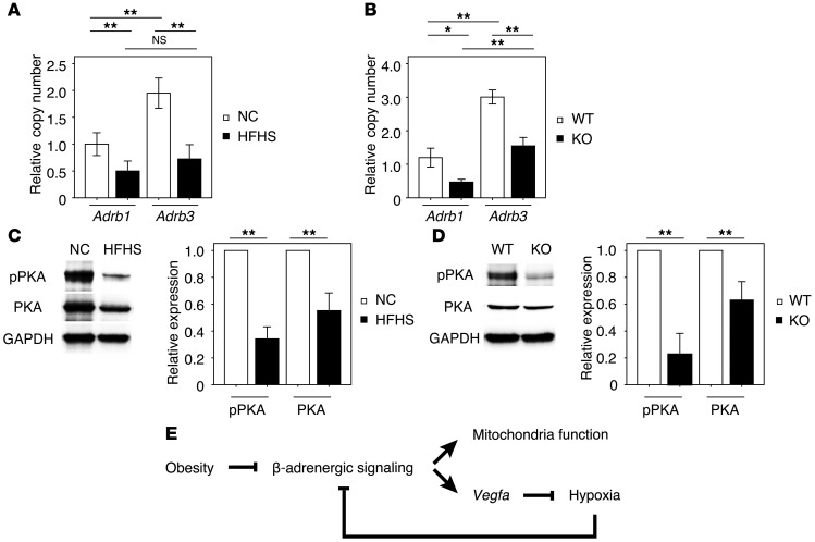
Brown adipose tissue (BAT) is a highly vascularized organ with abundant mitochondria that produce heat through uncoupled respiration. Obesity is associated with a reduction of BAT function; however, it is unknown how obesity promotes dysfunctional BAT. Here, using a murine model of diet-induced obesity, we determined that obesity causes capillary rarefaction and functional hypoxia in BAT, leading to a BAT "whitening" phenotype that is characterized by mitochondrial dysfunction, lipid droplet accumulation, and decreased expression of Vegfa. Targeted deletion of Vegfa in adipose tissue of nonobese mice resulted in BAT whitening, supporting a role for decreased vascularity in obesity-associated BAT. Conversely, introduction of VEGF-A specifically into BAT of obese mice restored vascularity, ameliorated brown adipocyte dysfunction, and improved insulin sensitivity. The capillary rarefaction in BAT that was brought about by obesity or Vegfa ablation diminished β-adrenergic signaling, increased mitochondrial ROS production, and promoted mitophagy. These data indicate that overnutrition leads to the development of a hypoxic state in BAT, causing it to whiten through mitochondrial dysfunction and loss. Furthermore, these results link obesity-associated BAT whitening to impaired systemic glucose metabolism.


Figures

Figure 1
Figure 1. The whitening of BAT associated with capillary rarefaction in diet-induced obesity.
(A) H&E staining of BAT from mice fed NC or HFHS diet. Scale bar: 50 μm. Right graph shows the number of large lipid droplets/field in BAT (×400, n = 4). (B) Electron micrographs of BAT from mice fed NC or HFHS diet. Right graph shows the number of mitochondria/cell (n = 3). Scale bar: 10 μm. (C and D) Real-time PCR expression of the mitochondrial-encoded transcript ND5 and the nucleus-encoded transcripts Ucp1, Ndufa, Atp5a, and Ppargc1a in BAT from mice fed NC or HFHS diet (n = 3–6). (E) Acute CTT for mice fed NC or HFHS (n = 5–7). (F) Immunofluorescent staining to detect blood vessels with Fluorescein Griffonia (Bandeiraea) Simplicifolia Lectin I (green) and adipocytes with Bodipy-TR (red) in BAT and WAT from mice fed NC or HFHS diet. Scale bars: 100 μm. (G and H) Pimonidazole staining (G) and positive area (H) in BAT and WAT of mice fed NC or HFHS diet determined by hypoxyprobe-1 staining (n = 4–6). Scale bars: 50 μm. (I) Oxygen levels in adipose tissues (AT pO2 [mmHg]) (n = 5–6). (J) Real-time PCR expression of Vegfa and Kdr in BAT and WAT of mice fed NC or HFHS diet (n = 4–10). Data were analyzed by 2-tailed Student’s t test (AD and H) or ANOVA (E, I, and J). *P < 0.05; **P < 0.01. All values represent the mean ± SEM.
Figure 2
Figure 2. The whitening of BAT and impaired glucose metabolism in aP2-Cre+/– Vegfaflox/flox mice.
(A) Real-time PCR expression of Vegfa and Kdr in BAT and WAT of aP2-Cre+/– Vegfaflox/flox (KO) and control Vegfaflox/flox mice (WT) (n = 4–19). (B) Immunofluorescent staining to detect vasculature with Fluorescein Griffonia (Bandeiraea) Simplicifolia Lectin I (green) and adipocytes with Bodipy-TR (red) in BAT and WAT from WT and KO mice. Scale bars: 100 μm. (C) Pimonidazole staining of BAT and WAT from WT and KO mice was performed by the hypoxyprobe-1 method. Scale bars: 50 μm. (D) H&E staining of BAT and WAT from WT and KO mice. Scale bars: 50 μm. (E) Real-time PCR detecting expression of ND5, Ucp1, Ndufa, Atp5a, and Ppargc1a in WAT and BAT of WT and KO mice (n = 3–6). (F and G) Electron micrographs of BAT of WT and KO mice (F) and the number of mitochondria/cell (G) (n = 3). Scale bar: 10 μm. (H) Acute CTT for mice prepared in A (n = 7). (I) GTT and ITT of mice prepared in A (n = 5–7). Data were analyzed by 2-tailed Student’s t test (E and G) or ANOVA (A, H, and I). *P < 0.05; **P < 0.01. All values represent the mean ± SEM.
Figure 3
Figure 3. BAT-specific Vegfa delivery induces the rebrowning of the whitened BAT in dietary obesity.
(A and B) Real-time PCR analysis of Vegfa and Kdr expression in BAT (A) and WAT (B) of mice after injection of ad-vegfa or control vector into BAT of mice fed NC or HFHS diets. ad-lacZ was used as a control (Con) (n = 5–8). (C) Immunofluorescent staining with Fluorescein Griffonia (Bandeiraea) Simplicifolia Lectin I (green) to detect vasculature and with Bodipy-TR (red) to detect lipid in BAT from NC- or HFHS-fed mice after the injection of ad-vegfa of the control adenoviral vector. Scale bar: 100 μm. (D) H&E staining of BAT from NC- and HFHS-fed mice prepared in A after the injection of ad-vegfa or a control adenoviral vector. Scale bar: 50 μm. (E) Quantitative analysis of the number of large lipid droplets/field in BAT under the different experimental conditions (×400, n = 4). (F) Real-time PCR analysis of the expression of ND5, Ucp1, Ndufa, and Ppargc1a in BAT of mice described in A (n = 3–10). (G) Acute CTT of the different experimental groups of mice (n = 3–7). (H) Glucose uptake by BAT was evaluated by measuring 2DG uptake (n = 4–6). (I) GTT and ITT in the different experimental groups of mice receiving ad-vegfa or control adenovirus (n = 4–8). Data were analyzed by ANOVA (A, B, and EI). *P < 0.05; **P < 0.01. All values represent the mean ± SEM.
Figure 4
Figure 4. VEGF-A–mediated regulation of mitochondrial ROS production and autophagic responses in the BAT of obese mice.
(A and B) FACS analysis for mitochondrial ROS (MitoSOX; A) and membrane potential (MitoRed with or without CCCP treatment; B) using isolated mitochondria extracted from BAT. (C) Western blot analysis of LC3A/B expression in BAT. The right graph indicates the quantification of LC3A/B-II expression relative to GAPDH-loading control (n = 3). (D and F) Immunofluorescent staining showing mitochondrial membrane protein Tom20 (green) colocalizing with autophagosomal membrane protein LC3 (red) in BAT of mice fed NC or HFHS diet with (F) or without (D) the injection of ad-vegfa or a control adenoviral vector (Con). Representative photomicrograph observed at ×3000 magnification. Scale bar: 3 μm. Merged areas are indicated by white arrows. The graph at right quantifies the number of puncta double stained with Tom20 and LC3 measured on 10 random fields and observed at ×3000 magnification (n = 3). (E and G) Real-time PCR expression of Bnip3 and Map1lc3b in BAT of mice (n = 3–6). (HJ) Western blot analysis of PINK1 (H), Parkin (I), and ubiquitin-conjugated protein (J) expression in isolated mitochondria extracted from BAT of mice. The graphs at right indicate the quantification relative to the expression of the Cox IV loading control (n = 3). In J, the level of ubiquitination is compared with the 25 kDa protein between the groups. Data were analyzed by 2-tailed Student’s t test (CE and HJ) or ANOVA (F and G). *P < 0.05; **P < 0.01. All values represent the mean ± SEM.
Figure 5
Figure 5. Hypoxia inhibits β-adrenergic signaling.
(A) Relative copy number of Adrb1 and Adrb3 transcript expression in BAT under these experimental conditions (n = 4–5). (B) Relative copy number of Adrb1 and Adrb3 transcript expression in BAT of the KO and WT mice (n = 3–8). (C and D) Western blot analysis of phosphorylated PKA (pPKA Thr197) and total PKA in BAT from mice fed NC or HFHS diet or BAT from aP2-Cre+/– Vegfaflox/flox (KO) and control Vegfaflox/flox mice (WT). Right graphs indicate quantification relative to PKA (for pPKA) and GAPDH-loading control (for PKA) (n = 3). (E) Graphical illustration of the downregulation of adrenergic signaling under conditions of obesity and the proposed positive feedback loop caused by hypoxic conditions. Data were analyzed by 2-tailed Student’s t test (C and D) or ANOVA (A and B). *P < 0.05; **P < 0.01. All values represent the mean ± SEM.

References

    1. Chechi K, Carpentier AC, Richard D. Understanding the brown adipocyte as a contributor to energy homeostasis. Trends Endocrinol Metab. 2013;24(8):408–420. - PubMed
    1. Tseng YH, Cypess AM, Kahn CR. Cellular bioenergetics as a target for obesity therapy. Nat Rev Drug Discov. 2010;9(6):465–482. doi: 10.1038/nrd3138. - DOI - PMC - PubMed
    1. Seale P, Lazar MA. Brown fat in humans: turning up the heat on obesity. Diabetes. 2009;58(7):1482–1484. doi: 10.2337/db09-0622. - DOI - PMC - PubMed
    1. Cypess AM, et al. Identification and importance of brown adipose tissue in adult humans. N Engl J Med. 2009;360(15):1509–1517. doi: 10.1056/NEJMoa0810780. - DOI - PMC - PubMed
    1. Bartelt A, et al. Brown adipose tissue activity controls triglyceride clearance. Nat Med. 2011;17(2):200–205. doi: 10.1038/nm.2297. - DOI - PubMed

Publication types

MeSH terms

Substances