. 2014 May 30;289(22):15544-53.
doi: 10.1074/jbc.M113.507616. Epub 2014 Apr 8.

Staphylococcal SplB serine protease utilizes a novel molecular mechanism of activation

Affiliations

Staphylococcal SplB serine protease utilizes a novel molecular mechanism of activation

Katarzyna Pustelny et al. J Biol Chem. .

Abstract

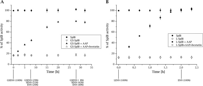
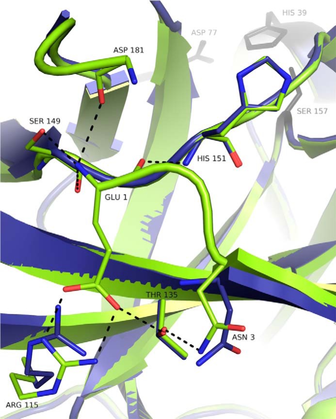
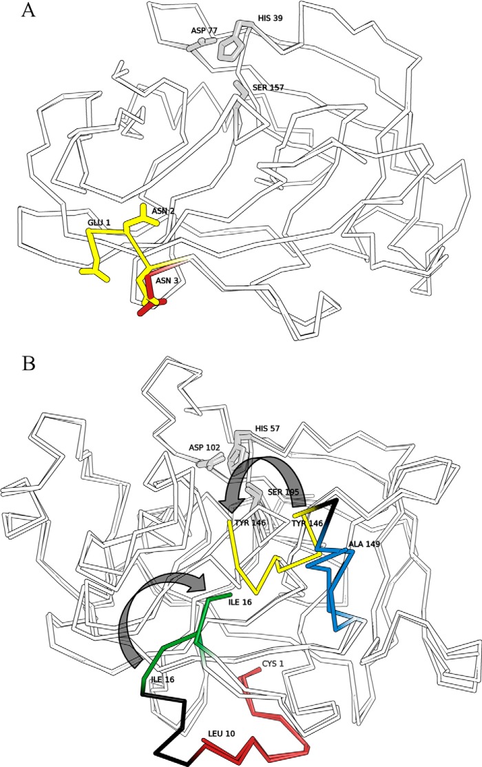
Staphylococcal SplB protease belongs to the chymotrypsin family. Chymotrypsin zymogen is activated by proteolytic processing at the N terminus, resulting in significant structural rearrangement at the active site. Here, we demonstrate that the molecular mechanism of SplB protease activation differs significantly and we characterize the novel mechanism in detail. Using peptide and protein substrates we show that the native signal peptide, or any N-terminal extension, has an inhibitory effect on SplB. Only precise N-terminal processing releases the full proteolytic activity of the wild type analogously to chymotrypsin. However, comparison of the crystal structures of mature SplB and a zymogen mimic show no rearrangement at the active site whatsoever. Instead, only the formation of a unique hydrogen bond network, distant form the active site, by the new N-terminal glutamic acid of mature SplB is observed. The importance of this network and influence of particular hydrogen bond interactions at the N terminus on the catalytic process is demonstrated by evaluating the kinetics of a series of mutants. The results allow us to propose a consistent model where changes in the overall protein dynamics rather than structural rearrangement of the active site are involved in the activation process.

Keywords: Crystal Structure; Enzyme Inactivation; Protease; Serine Protease; Signal Peptide; Staphylococcus aureus; Zymogen Activation.


Figures

FIGURE 1.
FIGURE 1.
Removal of artificial N-terminal extension releases the proteolytic activity of GS-SplB and l-SplB. A, GS-SplB was incubated with APP, and the proteolytic activity against Ac-WELQ-ACC (specific for SplB) was monitored. The activity is represented as a percentage of the maximum activity observed for the same concentration of SplB protease as the initial concentration of GS-SplB. Bestatin inhibits AAP activity. Samples were collected at indicated time points and subjected to N-terminal Edman degradation sequencing (results shown at the bottom of the panel). Activity of AAP remained unchanged during the experiment as monitored using Leu-pNa (not shown). B, same as in A but for L-SplB.
FIGURE 2.
FIGURE 2.
Hydrogen bond network involving N-terminal residues of mature SplB protease is absent in GS-SplB precursor mimic. Activation of SplB protease precursor mimic (blue, GS-SplB; PDB code 4K1S) by proteolytic processing is associated with formation of an extended hydrogen bond network at the newly formed N terminus of mature SplB protease (green; PDB code 2VID). Residues Gly-(−2) through Asn-2 of the precursor mimic are not defined by electron density and therefore not present in the model. Hydrogen bonds in SplB are indicated as black dotted lines. Water molecules are not shown for clarity.
FIGURE 3.
FIGURE 3.
Activation of SplB protease takes place without broad structural rearrangement in the vicinity of the active site contrary to the activation of chymotrypsinogen. Overlays of active and zymogen forms of serine proteases are shown. Regions of pronounced structural rearrangements are highlighted in color. Catalytic triad residues are highlighted gray. A, overlay of GS-SplB (PDB code 4K1S) and SplB (PDB code 2VID) crystal structures. N-terminal residues are depicted in red and yellow, respectively. Gly-(−1)–Asn-2 are undefined by electron density in the structure of GS-SplB. B, overlay of chymotrypsinogen (PDB code 2CGA) and chymotrypsin (PDB code 1YPH) crystal structures. Regions removed during proteolytic activation are highlighted black. N-terminal peptide of the precursor (red) is processed by trypsin to release a new N terminus (green). Regions of the “activation domain” rearranged upon precursor activation are highlighted yellow and blue.

References

    1. Władyka B., Pustelny K. (2008) Regulation of bacterial protease activity. Cell. Mol. Biol. Lett. 13, 212–229 - PMC - PubMed
    1. López-Otín C., Bond J. S. (2008) Proteases: multifunctional enzymes in life and disease. J. Biol. Chem. 283, 30433–30437 - PMC - PubMed
    1. Strausberg S., Alexander P., Wang L., Schwarz F., Bryan P. (1993) Catalysis of a protein folding reaction: thermodynamic and kinetic analysis of subtilisin BPN′ interactions with its propeptide fragment. Biochemistry 32, 8112–8119 - PubMed
    1. Baker D., Shiau A. K., Agard D. A. (1993) The role of pro regions in protein folding. Curr. Op. Cell Biol. 5, 966–970 - PubMed
    1. Winther J. R., Sørensen P. (1991) Propeptide of carboxypeptidase Y provides a chaperon-like functions as well as inhibition of enzymatic activity. Proc. Natl. Acad. Sci. U.S.A. 88, 9330–9334 - PMC - PubMed

Publication types

MeSH terms

LinkOut - more resources