. 2014 Apr 8;9(4):e93809.
doi: 10.1371/journal.pone.0093809. eCollection 2014.

Biochemical properties of ectoine hydroxylases from extremophiles and their wider taxonomic distribution among microorganisms

Affiliations

Biochemical properties of ectoine hydroxylases from extremophiles and their wider taxonomic distribution among microorganisms

Nils Widderich et al. PLoS One. .

Abstract

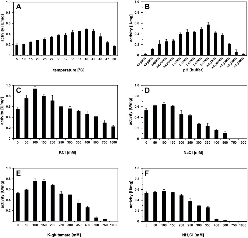
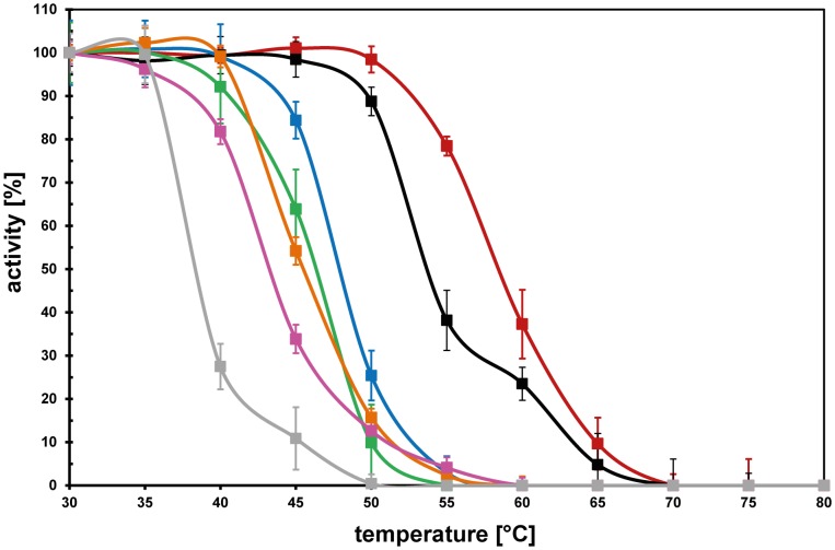
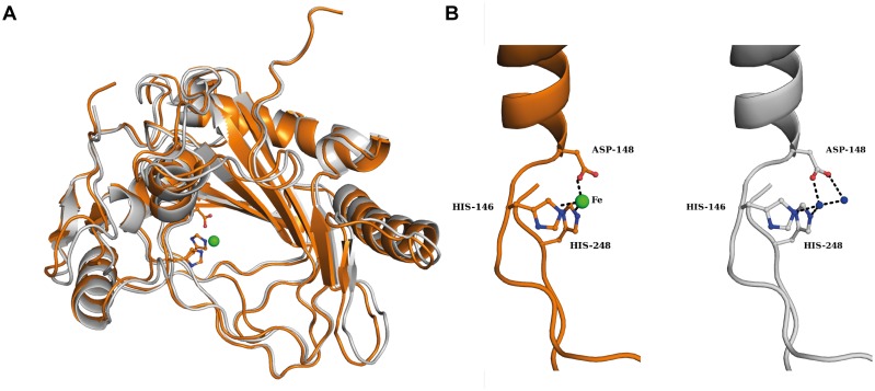
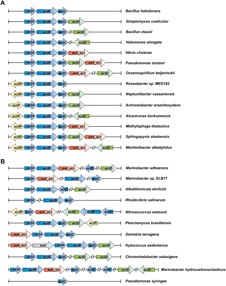
Ectoine and hydroxyectoine are well-recognized members of the compatible solutes and are widely employed by microorganisms as osmostress protectants. The EctABC enzymes catalyze the synthesis of ectoine from the precursor L-aspartate-β-semialdehyde. A subgroup of the ectoine producers can convert ectoine into 5-hydroxyectoine through a region-selective and stereospecific hydroxylation reaction. This compatible solute possesses stress-protective and function-preserving properties different from those of ectoine. Hydroxylation of ectoine is carried out by the EctD protein, a member of the non-heme-containing iron (II) and 2-oxoglutarate-dependent dioxygenase superfamily. We used the signature enzymes for ectoine (EctC) and hydroxyectoine (EctD) synthesis in database searches to assess the taxonomic distribution of potential ectoine and hydroxyectoine producers. Among 6428 microbial genomes inspected, 440 species are predicted to produce ectoine and of these, 272 are predicted to synthesize hydroxyectoine as well. Ectoine and hydroxyectoine genes are found almost exclusively in Bacteria. The genome context of the ect genes was explored to identify proteins that are functionally associated with the synthesis of ectoines; the specialized aspartokinase Ask_Ect and the regulatory protein EctR. This comprehensive in silico analysis was coupled with the biochemical characterization of ectoine hydroxylases from microorganisms that can colonize habitats with extremes in salinity (Halomonas elongata), pH (Alkalilimnicola ehrlichii, Acidiphilium cryptum), or temperature (Sphingopyxis alaskensis, Paenibacillus lautus) or that produce hydroxyectoine very efficiently over ectoine (Pseudomonas stutzeri). These six ectoine hydroxylases all possess similar kinetic parameters for their substrates but exhibit different temperature stabilities and differ in their tolerance to salts. We also report the crystal structure of the Virgibacillus salexigens EctD protein in its apo-form, thereby revealing that the iron-free structure exists already in a pre-set configuration to incorporate the iron catalyst. Collectively, our work defines the taxonomic distribution and salient biochemical properties of the ectoine hydroxylase protein family and contributes to the understanding of its structure.


Conflict of interest statement

Competing Interests: The authors have declared that no competing interests exist.

Figures

Figure 1
Figure 1. Phylogenetic tree of EctC- and EctD-type proteins.
The shown phylogenetic tree is based on the alignment of EctC amino acid sequences identified by a BLAST search at the JGI Web-server that were then aligned using ClustalW. These compiled amino acid sequences were then used to assess the phylogenetic distribution of the EctC protein using the iTOL Web-server. Evolutionary distances are not given. The color code indicates the distribution of EctC among members of the Bacteria and Archaea. The presence of an ectD gene in a given microbial species possessing ectC is indicated by black (ectD is part of the ect gene cluster) or red circles (ectD is located outside of the ect gene cluster). Purple circles are indicating the presence of an ask_ect gene associated with the ect gene cluster, whereas the presence of an ectR regulatory gene is indicted by green circles. If different strains of the same species were sequenced, only one representative symbolizes them. For instance, there are genomic data of 139 strain of Vibrio cholerae available in the database, each of which possesses an ectABC gene cluster, but only one of these sequences was used for the phylogenetic analysis.
Figure 2
Figure 2. Purification of recombinant EctD proteins.
A 12% SDS-PAGE of the recombinant EctD-type proteins originating from different microbial species after their overproduction in E. coli and purification via Strep-tag-II affinity chromatography is shown. 5 μg of each purified EctD protein were applied onto the gel. The gel was run at 25 mA for 2.5 h. The PageRuler Prestained Protein Ladder (Thermo Scientific, Schwerte, Germany) was used as marker. We note the presence of a overlapping second band in protein sample of the Paenibacillus lautus EctD preparation. This might stem from overloading the gel somewhat or from partial degradation of the purified EctD protein.
Figure 3
Figure 3. Biochemical properties of the EctD enzyme from S. alaskensis.
The enzyme activity of the ectoine hydroxylase from S. alaskensis is shown with respect to (A) the temperature optimum, (B) the pH optimum and the influence of different salts: (C) potassium chloride, (D) sodium chloride, (E) potassium glutamate and (F) ammonium chloride.
Figure 4
Figure 4. Resistance of various ectoine hydroxylases against the denaturing effects of high temperature.
The temperature profiles of the ectoine hydroxylases from H. elongata (grey), A. cryptum (pink), A. ehrlichii (orange), V. salexigens (green), P. stutzeri (blue), S. alaskensis (black), and P. lautus (red) are given. Each EctD protein was pre-incubated at the indicated temperatures for 15 min before its specific activity was then determined under its optimal assay condition. The enzyme activity exhibited by each enzyme after pre-incubation at 30°C was set as 100%.
Figure 5
Figure 5. Crystal structure of the apo-form of the ectoine hydroxylase from V. salexigens.
(A) Overlay of the crystal structure of the apo-EctD protein (colored in grey) with the Fe-bound crystal structure of EctD (colored in orange) in cartoon representation. The Fe ion of the Fe-bound EctD protein is represented as a green sphere. Data coordinates for the iron-bound form of the V. salexigens EctD protein were taken from the protein database (PDB) entry 3EMR and those from the iron-free form were from PDB entry 4NMI. (B) Details of the molecular determinants of the iron-binding site of the V. salexigens EctD protein in its iron bound (orange) and iron-free (grey) forms. The side chains of the iron-binding residues Asp148, His146 and His248 are highlighted. Green and blue spheres represent the bound iron and water molecules, respectively.
Figure 6
Figure 6. Genetic organization of the ectoine/hydroxyectoine biosynthesis gene clusters.
The different types of ect gene clusters present in putative ectoine/hydroxyectoine producers are represented. An example for the genetic organization of each type of ect cluster found in the ectC reference set (440 representatives; Fig. 1) is given along with a microorganism in which it occurs. (A) Most common organizational types of the ect gene clusters. (B) Representatives of the organization of the ectoine/hydroxyectoine biosynthetic genes that deviate from the otherwise commonly found genetic organization.

References

    1. Bremer E, Krämer R (2000) Coping with osmotic challenges: osmoregulation through accumulation and release of compatible solutes. In: Storz G, Hengge-Aronis R, editors. Bacterial Stress Responses: ASM Press. 79–97.
    1. Wood JM, Bremer E, Csonka LN, Krämer R, Poolman B, et al. (2001) Osmosensing and osmoregulatory compatible solute accumulation by bacteria. Comp Biochem Physiol Part A Mol Integr Physiol 130: 437–460. - PubMed
    1. da Costa MS, Santos H, Galinski EA (1998) An overview of the role and diversity of compatible solutes in Bacteria and Archaea. Adv Biochem Eng Biotechnol 61: 117–153. - PubMed
    1. Kempf B, Bremer E (1998) Uptake and synthesis of compatible solutes as microbial stress responses to high osmolality environments. Arch Microbiol 170: 319–330. - PubMed
    1. Roeβler M, Müller V (2001) Osmoadaptation in bacteria and archaea: common principles and differences. Env Microbiol Rep 3: 743–754. - PubMed

Publication types

Associated data