. 2014 Jun;34(6):1082-8.
doi: 10.1038/jcbfm.2014.62. Epub 2014 Apr 9.

Brain sparing in fetal mice: BOLD MRI and Doppler ultrasound show blood redistribution during hypoxia

Affiliations

Brain sparing in fetal mice: BOLD MRI and Doppler ultrasound show blood redistribution during hypoxia

Lindsay S Cahill et al. J Cereb Blood Flow Metab. 2014 Jun.

Abstract

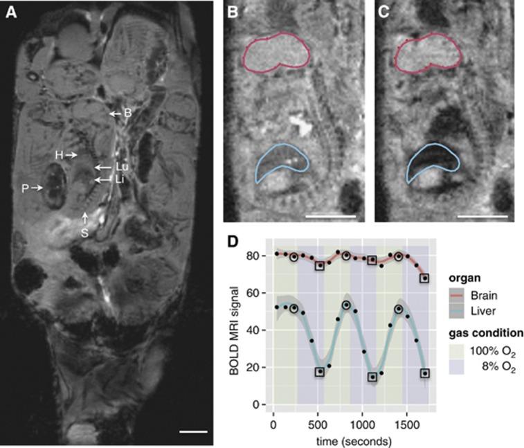
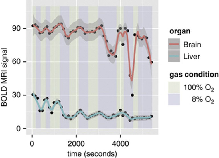
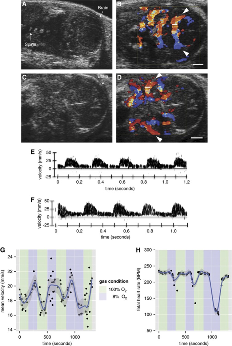
Mice reproduce many features of human pregnancy and have been widely used to model disorders of pregnancy. However, it has not been known whether fetal mice reproduce the physiologic response to hypoxia known as brain sparing, where blood flow is redistributed to preserve oxygenation of the brain at the expense of other fetal organs. In the present study, blood oxygen level-dependent (BOLD) magnetic resonance imaging (MRI) and Doppler ultrasound were used to determine the effect of acute hypoxia on the fetal blood flow in healthy, pregnant mice. As the maternal inspired gas mixture was varied between 100% and 8% oxygen on the timescale of minutes, the BOLD signal intensity decreased by 44±18% in the fetal liver and by 12±7% in the fetal brain. Using Doppler ultrasound measurements, mean cerebral blood velocity was observed to rise by 15±8% under hypoxic conditions relative to hyperoxia. These findings are consistent with active regulation of cerebral oxygenation and clearly show brain sparing in fetal mice.


Figures

Figure 1
Figure 1
Maternal physiologic changes under repeated exposure to acute hypoxia. Scatter plots showing the time course of (A) maternal arterial blood oxygen saturation (%) and (B) maternal heart rate (beats per minute, BPM) as the inspired oxygen mixture is varied. The spikes in the maternal heart rate at 76 and 387 seconds are attributed to instrument noise.
Figure 2
Figure 2
Fetal mouse blood oxygen level-dependent contrast magnetic resonance imaging (MRI) signal variation with maternal inspired oxygen. (A) Representative coronal anatomic MR image of a 17.5-day gestation dam. Multiple fetuses and placentas are seen in this view. B: brain; H: heart; Li: liver; Lu: lung; P: placenta; S: spine. (B) Representative blood oxygen level-dependent (BOLD) MR image of a fetus showing the placement of regions of interest (ROIs) when the dam is breathing 100% O2 and (C) 8% O2. The decrease in signal intensity (SI) in the liver during hypoxia is clearly visible. (D) Scatter plot showing the absolute BOLD MR signal (arbitrary units) in one of the six dams (B, C) as the inspired oxygen mixture is varied. The smoothing (solid red and blue lines) was performed using LOWESS, a robust locally weighted smoothing algorithm. SIhypoxia and SIhyperoxia were determined by calculating the mean of the SI at the final time point of each gas cycle under 8% O2 (squares) and 100% O2 (circles), respectively. Scale bars in (A), (B), and (C)=5 mm.
Figure 3
Figure 3
Fetal mouse blood oxygen level-dependent contrast magnetic resonance imaging (MRI) signal variation with prolonged hypoxic challenge shows the limits of the brain sparing mechanism. Scatter plot showing the absolute blood oxygen level-dependent (BOLD) MR signal (arbitrary units) in one of the six dams as the inspired oxygen mixture is varied. The smoothing (solid red and blue lines) was performed using LOWESS.
Figure 4
Figure 4
Assessment of mouse fetal blood flow by ultrasound biomicroscopy. (A, C) Representative B-mode images of E17.5 fetuses in coronal section showing the fetal brain. Color Doppler overlay indicating flow direction (red is blood flowing toward the transducer and blue is blood flowing away from the transducer) for the (B) posterior cerebral arteries (PCAs) (arrows shown) and (D) middle cerebral arteries (MCAs) (arrows shown). The angle of insonation was 0° and 5° for (B) and (D), respectively. Measurements were taken from the vessel in the near field with the transducer located above the structure in (B) and (D). The white bars on the vessel of interest represent the location of the Doppler sample volume. (E) Doppler blood velocity spectrum versus time for the PCA when the dam is breathing 100% O2 and (F) 8% O2. Similar waveforms were recorded for the MCA. (G) Scatter plot showing the mean PCA blood flow as the inspired oxygen mixture is varied. (H) Scatter plot showing the fetal heart rate (beats per minute, BPM) as the inspired oxygen mixture is varied. The smoothing (blue line) was performed using LOWESS. Scale bars in (B) and (D)=1 mm.

References

    1. Arraut AM, Frias AE, Hobbs TR, McEvoy C, Spindel ER, Rasanen J. Fetal pulmonary arterial vascular impedance reflects changes in fetal oxygenation at near-term gestation in a nonhuman primate model. Reprod Sci. 2013;20:33–38. - PMC - PubMed
    1. Hill A. Current concepts of hypoxic-ischemic cerebral injury in the term newborn. Pediatr Neurol. 1991;7:317–325. - PubMed
    1. Lou HC. Perinatal hypoxic-ischemic brain damage and intraventricular hemorrhage. A pathogenetic model. Arch Neurol. 1980;37:585–587. - PubMed
    1. Cohn HE, Sacks EJ, Heymann MA, Rudolph AM. Cardiovascular responses to hypoxemia and acidemia in fetal lambs. Am J Obstet Gynecol. 1974;120:817–824. - PubMed
    1. Gleason CA, Hamm C, Jones MD., Jr. Effect of acute hypoxemia on brain blood flow and oxygen metabolism in immature fetal sheep. Am J Physiol. 1990;258:H1064–H1069. - PubMed

Publication types

LinkOut - more resources