Review
. 2014;8(3):191-204.
doi: 10.4161/cam.27840.

Podosome organization drives osteoclast-mediated bone resorption

Affiliations
Review

Podosome organization drives osteoclast-mediated bone resorption

Dan Georgess et al. Cell Adh Migr. 2014.

Abstract

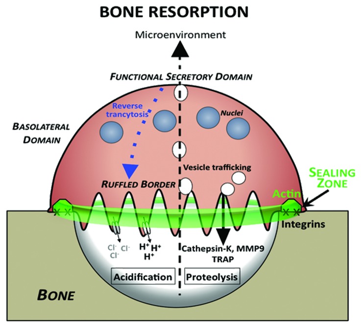
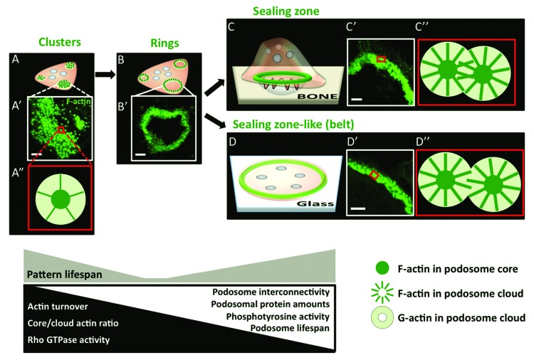
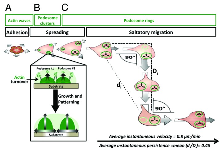
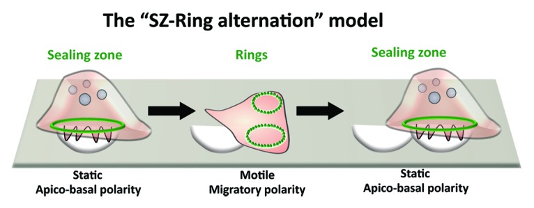
Osteoclasts are the cells responsible for physiological bone resorption. A specific organization of their most prominent cytoskeletal structures, podosomes, is crucial for the degradation of mineralized bone matrix. Each podosome is constituted of an F-actin-enriched central core surrounded by a loose F-actin network, called the podosome cloud. In addition to intrinsic actin dynamics, podosomes are defined by their adhesion to the extracellular matrix, mainly via core-linking CD44 and cloud-linking integrins. These properties allow podosomes to collectively evolve into different patterns implicated in migration and bone resorption. Indeed, to resorb bone, osteoclasts polarize, actively secrete protons, and proteases into the resorption pit where these molecules are confined by a podosome-containing sealing zone. Here, we review recent advancements on podosome structure and regulatory pathways in osteoclasts. We also discuss the distinct functions of different podosome patterns during the lifespan of a single osteoclast.

Keywords: actin; actin rings; bone degradation; migration; osteoclasts; podosomes; sealing zone.


Figures

None
Figure 1. Bone-resorbing osteoclasts are polarized. In contact with bone, OCs exhibit functional membrane domains defining apico-basal polarity. At the bone surface level they form the actin-rich sealing zone (green) around integrins, such as αVβ3. Sealing zone delineates the ruffled border (RB) where bone degradation takes place. The ruffled border is formed as a consequence of trafficking of vesicles in the endosomal pathway, and therefore, has characteristics of a late endosomal membrane. H+ protons are generated through the activity of carbonic anhydrase II and excreted in the resorption lacuna through V-ATPase, leading to acidification and mineral dissolution. In addition, proteases are secreted in the lacuna through the ruffled border leading to degradation of organic matrix components. Simultaneously, degradation products from the resorption process are removed from the resorption lacuna by a transcytotic pathway and released at the functional secretory domain (FSD). A reverse pathway from the functional secretion domain to the ruffled border has been identified (blue).
None
Figure 2. Current model of dynamic podosome patterning in OCs. Podosomes (green) are collectively arranged into clusters (A), rings (B), and then finally into either sealing zones (SZ) on glass (C) or SZ-like structures (also called “belts”) on non-mineralized substrates (D). In the cluster, podosomes are grouped in close vicinity as can be seen by immunofluorescence (IF) in an area of the cell (A’) and have a “relaxed” architecture (A”). Podosome clusters evolve to transient ring patterns. The SZ ensures proper bone resorption by confining the degradation molecules secreted by the OC ruffled border into the resorption lacuna. The SZ-like structure is not associated with a functional activity and is at the extreme periphery of the cell. There is an increase in podosome density and interconnectivity in the SZ (C”) and the SZL (D”) compared with the cluster. Kinetic, biochemical, and structural properties that accompany the patterning process are also displayed in this scheme. Scale bar in all micrographs is 5 µm.
None
Figure 3. Osteoclast adhesion, spreading, and migration. (A) When the osteoclasts first adheres to its substrate, it is contracted and radial waves of disorganized actin are consistent with the initiation of its spreading. (B) At the early stages of spreading, podosomes form at the periphery of the osteoclast and cluster at the edges of the cell. The dynamic turnover of actin within each podosome leads to podosome growth. Growing neighboring podosomes apply forces on one another and, to the substrate if possible, thus leading to further spreading of the cell periphery. Individual podosome growth during osteoclast spreading also leads to their collective patterning from clusters into circular superstructures called podosome rings. (C) Podosome rings continue to display the actin dynamics and podosome growth observed in clusters. The expansion of individual rings generates forces that drive osteoclast displacement. A typical mode of osteoclast motility on two-dimensional substrates, called saltatory migration, is a biphasic cycle, namely due to ring assembly and disassembly in different areas of the cell. In the first phase, podosome rings expanding at the same side of the cell, drive osteoclast displacement in a straightforward direction. The second phase is consistent with the disassembly/collapse of one or more rings and the continued expansion of the remaining ring leads to a 90 °C-angular turn in direction. Soon after the angular turn, new rings are formed and determine the new leading edge of the osteoclast, thus restarting a second phase of straightforward movement. The latter ends with a new angular turn and the biphasic cycle of migration is therefore maintained for several rounds, allowing the osteoclast to cover a wide area of the substrate. Saltatory osteoclast migration can be characterized by quantifiable parameters, such as an average velocity of 0.8 µm/min and an average instantaneous persistence of 0.45. This mode of migration as well as its quantified parameters have been characterized in osteoclasts migration on culture-treated dishes.
None
Figure 4. Osteoclast migration and resorption. Typically, osteoclasts resorb bone by forming resorption pit trails. In the current model for bone degradation and migration (SZ-Ring alternation), bone-resorbing osteoclasts are apico-basally polarized with a sealing zone. When resorption stops, they flatten and start migration by becoming polarized with a leading and a trailing edge. Then, osteoclast will stop again and start a new cycle of resorption.

References

    1. Bartl R. Osteoporosis: diagnosis, prevention, therapy. New York: Springer, 2009.
    1. Harre U, Schett G. Bone research in 2012: the ups and downs of bone in health and rheumatic disease. Nat Rev Rheumatol. 2013;9:67–8. doi: 10.1038/nrrheum.2012.219. - DOI - PubMed
    1. Harre U, Georgess D, Bang H, Bozec A, Axmann R, Ossipova E, Jakobsson PJ, Baum W, Nimmerjahn F, Szarka E, et al. Induction of osteoclastogenesis and bone loss by human autoantibodies against citrullinated vimentin. J Clin Invest. 2012;122:1791–802. doi: 10.1172/JCI60975. - DOI - PMC - PubMed
    1. McInnes IB, Schett G. The pathogenesis of rheumatoid arthritis. N Engl J Med. 2011;365:2205–19. doi: 10.1056/NEJMra1004965. - DOI - PubMed
    1. Lazner F, Gowen M, Pavasovic D, Kola I. Osteopetrosis and osteoporosis: two sides of the same coin. Hum Mol Genet. 1999;8:1839–46. doi: 10.1093/hmg/8.10.1839. - DOI - PubMed

Publication types

LinkOut - more resources