

Association of PD-1, PD-1 ligands, and other features of the tumor immune microenvironment with response to anti-PD-1 therapy
- PMID: 24714771
- PMCID: PMC4185001
- DOI: 10.1158/1078-0432.CCR-13-3271
Association of PD-1, PD-1 ligands, and other features of the tumor immune microenvironment with response to anti-PD-1 therapy
Abstract
Purpose: Immunomodulatory drugs differ in mechanism-of-action from directly cytotoxic cancer therapies. Identifying factors predicting clinical response could guide patient selection and therapeutic optimization.
Experimental design: Patients (N = 41) with melanoma, non-small cell lung carcinoma (NSCLC), renal cell carcinoma (RCC), colorectal carcinoma, or castration-resistant prostate cancer were treated on an early-phase trial of anti-PD-1 (nivolumab) at one institution and had evaluable pretreatment tumor specimens. Immunoarchitectural features, including PD-1, PD-L1, and PD-L2 expression, patterns of immune cell infiltration, and lymphocyte subpopulations, were assessed for interrelationships and potential correlations with clinical outcomes.
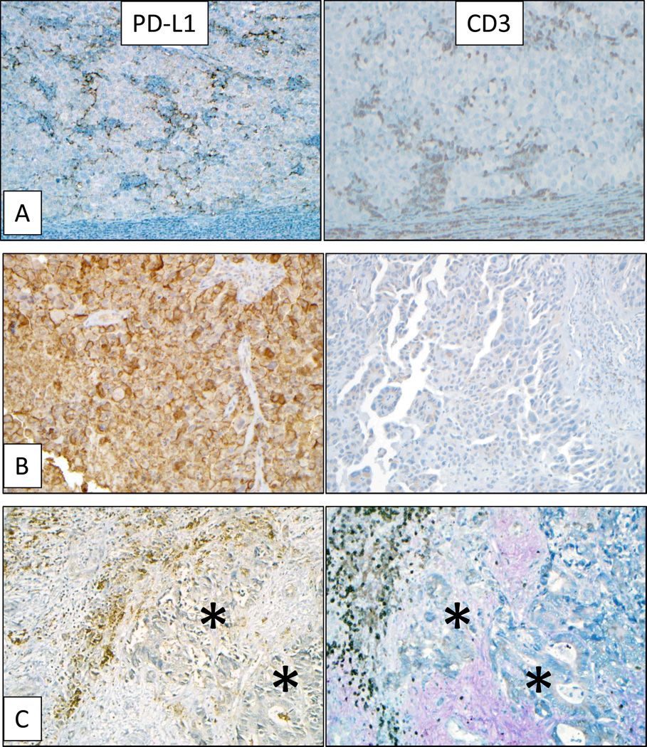
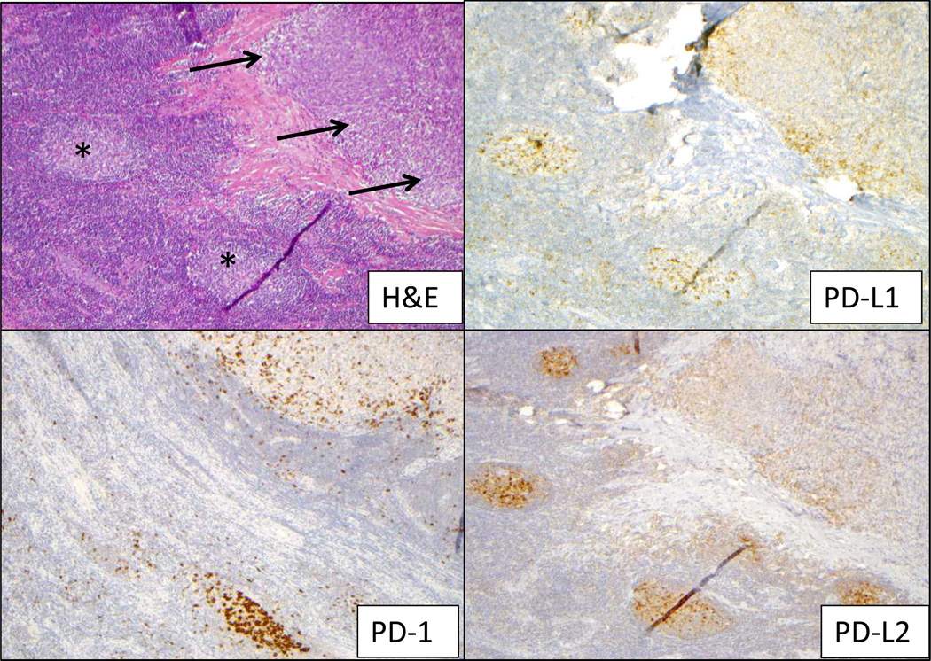
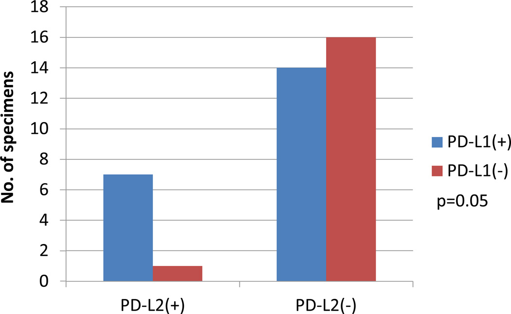
Results: Membranous (cell surface) PD-L1 expression by tumor cells and immune infiltrates varied significantly by tumor type and was most abundant in melanoma, NSCLC, and RCC. In the overall cohort, PD-L1 expression was geographically associated with infiltrating immune cells (P < 0.001), although lymphocyte-rich regions were not always associated with PD-L1 expression. Expression of PD-L1 by tumor cells and immune infiltrates was significantly associated with expression of PD-1 on lymphocytes. PD-L2, the second ligand for PD-1, was associated with PD-L1 expression. Tumor cell PD-L1 expression correlated with objective response to anti-PD-1 therapy, when analyzing either the specimen obtained closest to therapy or the highest scoring sample among multiple biopsies from individual patients. These correlations were stronger than borderline associations of PD-1 expression or the presence of intratumoral immune cell infiltrates with response.
Conclusions: Tumor PD-L1 expression reflects an immune-active microenvironment and, while associated other immunosuppressive molecules, including PD-1 and PD-L2, is the single factor most closely correlated with response to anti-PD-1 blockade. Clin Cancer Res; 20(19); 5064-74. ©2014 AACR.
©2014 American Association for Cancer Research.
Figures
References
-
- Agata Y, Kawasaki A, Nishimura H, Ishida Y, Tsubata T, Yagita H, et al. Expression of the PD-1 antigen on the surface of stimulated mouse T and B lymphocytes. Int Immunol. 1996;8:765–772. - PubMed
-
- Dong H, Zhu G, Tamada K, Chen L. B7-H1, a third member of the B7 family, co-stimulates T-cell proliferation and interleukin-10 secretion. Nat Med. 1999;5:1365–1369. - PubMed
-
- Dong H, Strome SE, Salomao DR, Tamura H, Hirano F, Flies DB, et al. Tumor-associated B7-H1 promotes T-cell apoptosis: A potential mechanism of immune evasion. Nat Med. 2002;8:793–800. - PubMed
-
- Mazanet MM, Hughes CC. B7-H1 is expressed by human endothelial cells and suppresses T cell cytokine synthesis. J Immunol. 2002;169:3581–3588. - PubMed
Publication types
MeSH terms
Substances
Grants and funding
LinkOut - more resources
Full Text Sources
Other Literature Sources
Medical
Research Materials
