Review
. 2014 Aug;71(16):3119-37.
doi: 10.1007/s00018-014-1609-0. Epub 2014 Apr 9.

Gene activation and cell fate control in plants: a chromatin perspective

Affiliations
Review

Gene activation and cell fate control in plants: a chromatin perspective

Julia Engelhorn et al. Cell Mol Life Sci. 2014 Aug.

Abstract

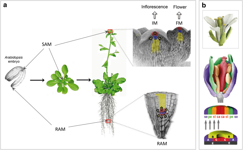
In plants, environment-adaptable organogenesis extends throughout the lifespan, and iterative development requires repetitive rounds of activation and repression of several sets of genes. Eukaryotic genome compaction into chromatin forms a physical barrier for transcription; therefore, induction of gene expression requires alteration in chromatin structure. One of the present great challenges in molecular and developmental biology is to understand how chromatin is brought from a repressive to permissive state on specific loci and in a very specific cluster of cells, as well as how this state is further maintained and propagated through time and cell division in a cell lineage. In this review, we report recent discoveries implementing our knowledge on chromatin dynamics that modulate developmental gene expression. We also discuss how new data sets highlight plant specificities, likely reflecting requirement for a highly dynamic chromatin.


Figures

Fig. 1
Fig. 1
Plants continuously produce organs from meristems maintained at their growing extremities (example of the Arabidopsis model plant). a Plant organogenesis occurs after embryogenesis, and most developmental programs (vegetative development, floral transition, flower organogenesis) are induced after seed germination. Continuous organogenesis is possible thanks to the maintenance of active populations of unspecialized, dividing stem cells at the growing apices. In the embryo, these stem cells are located at the shoot and root apical meristems (SAM and RAM, respectively). The RAM will produce all cells for the formation of primary and lateral roots, while the SAM will give rise to all aerial organs. Three-dimensional reconstructions of confocal views of RAM and SAM show, in blue, the organizing center, place of active cell divisions and source of stem cells and in yellow and red the cells that will enter differentiation, for formation of the stem tip, epidermis tissues and lateral organs. The yellow-filled arrows in the 3D reconstructions of confocal views show the direction of cell divisions. Upon germination, post-embryonic organogenesis proceeds from the very simple embryo structure: two embryonic leaves (cotyledons) expand, and the shoot apex starts to initiate leaves as first lateral organs. After several weeks of vegetative growth, in response to endogenous and environmental cues, the plant undergoes the transition to flowering. At this time, the SAM transits from a vegetative to a reproductive structure, the stem elongates and lateral branches and flowers are produced. After floral transition, the SAM is also called inflorescence meristem (IM) because it produces flower meristems (FM) on its flanks. b Each FM produces concentric whorls of organs that make a flower. The type of lateral organ that emerges from all meristematic structures (vegetative, reproductive, floral) highly depends on the specific expression of developmental genes in their founder, differentiating cells. In the case of the FM, the combinatorial expression patterns of specific genes (encoding MADS domain-containing transcription factors, described in the ABCE model) will lead to the formation of drastically different organs such as sepals (Se), petals (Pe), stamens (St) and carpels (Ca). [3D reconstructions from confocal images of SAM and RAM shown in (a) were reproduced from the Current Opinion in Plant Biology journal (Sablowski, 2011, published by Elsevier). Copyright for colorized scanning electron microscopy images of flowers shown in (b): J. Berger and D. Weigel, Max-Planck-Institute for Developmental Biology]
Fig. 2
Fig. 2
Polycomb and trithorax complexes targeting genes specific for the different developmental processes during the Arabidopsis life cycle. The inset shows the antagonistic functions of the Polycomb (PcG, PRC1 and PRC2 complexes) and the trithorax (trxG) complexes on target developmental gene (dev locus) expression. Three distinct PRC2, H3K27me3-depositing complexes exist in Arabidopsis: The FIS2 complex [composed of FIS2, MSI1, FERTILISATION-INDEPENDENT ENDOSPERM (FIE) and MEA] represses genes involved in gametogenesis and seed development (PHE1, FUS3, MEA). The VRN2 complex (composed of VRN2, MSI1, FIE and CLF or SWN) represses FLC, which is involved in the floral transition. The EMF2 complex (composed of EMF2, FIE, CLF or SWN and a MSI1 homolog) represses among others the floral MADS genes such as AG, AP3 and AGAMOUS-LIKE 19 (AGL19). LHP1 is proposed to recruit PRC1 complexes to H3K27me3-marked chromatin, which establishes stable gene repression. Components of PRC1 complexes are the RING domain containing proteins AtRING1a, AtRING1b and AtBMI1a-c, which function in ubiquitination of H2A. EMF1 is another putative PRC1 component in Arabidopsis, which was also shown to function independently of PRC1. The trxG ATX1, SDG2 and ULT1 proteins activate expression of genes involved in floral transition, flower organogenesis and gametogenesis (via direct deposition of H3K4me3 for ATX1 and SDG2). SDG2 and PKL were also shown to regulate root development. SYD, BRM and PKL are ATP-dependent chromatin remodelers shown to be involved in the activation of target developmental genes for most developmental processes. Phase transitions: G (germination), B (bolting), F (fertilization) and D (desiccation). [Schematic representation of the PRC2 complex was inspired by Ciferri et al. eLife 2012; 1:e00005]
Fig. 3
Fig. 3
Molecular events bringing a locus from an H3K27me3-repressed state to an H3K4me3-active state. Transcriptional status of the locus is indicated on the left hand side for each step. At the time of activation, transcriptional activators and transcription factors (TF) would recruit the REF6 H3K27-specific demethylase at a PcG-maintained repressed target locus (CLF is the HMT for H3K27; PRC2 is displayed in red), thus inducing removal of the repressive marks. The locus becomes competent for activation and likely carries the H3K4me2 mark (deposited by ATX2). At the time of pre-initiation complex (PIC) formation, the ULT1 protein may be instrumental in recruiting ATX1, which would stabilize the PIC via interaction with the non-phosphorylated form of the C-Terminal Domain (CTD) of Pol II and the TATA box binding protein at the promoter of target genes. Transcription is initiated and, at the early elongation step (Pol II has moved to the +300-bp region of the transcribed gene), CTD of Pol II, then Ser5 phosphorylated, recruits ATX1 to further facilitate trimethylation of H3K4 within the first 300-bp region of the transcribed gene. The BRM and SYD SWI/SNF2 ATPases may be recruited by TFs for nucleosome disassembly allowing accessibility of the target region to TFs and the general transcriptional machinery. Finally, ATX1-dependent deposition of me3 on H3K4 is required for further transcriptional elongation. Relative chronology between the above-described events has not been resolved yet, and it is unknown, for instance, whether repressive marks are all removed at the time of gene expression and whether active marks are already present at the locus while elongation takes place. The gray box includes the events of activation

Similar articles

Cited by

References

    1. Fornara F., Montaigu A., Coupland G. SnapShot: Control of flowering in Arabidopsis. Cell. 2010;141:550. - PubMed
    1. Kaufmann K, Nagasaki M, Jáuregui R. Modelling the molecular interactions in the flower developmental network of Arabidopsis thaliana. In Silico Biol. 2010;10:125–143. - PubMed
    1. Posé D, Yant L, Schmid M. The end of innocence: flowering networks explode in complexity. Curr Opin Plant Biol. 2012;15:45–50. - PubMed
    1. Coen ES, Meyerowitz EM. The war of the whorls: genetic interactions controlling flower development. Nature. 1991;353:31–37. - PubMed
    1. Krizek BA, Fletcher JC. Molecular mechanisms of flower development: an armchair guide. Nat Rev Genet. 2005;6:688–698. - PubMed

Publication types

LinkOut - more resources