Review
. 2015 Jan;73(1):102-16.
doi: 10.1002/mrm.25197. Epub 2014 Apr 8.

Recommended implementation of arterial spin-labeled perfusion MRI for clinical applications: A consensus of the ISMRM perfusion study group and the European consortium for ASL in dementia

Affiliations
Review

Recommended implementation of arterial spin-labeled perfusion MRI for clinical applications: A consensus of the ISMRM perfusion study group and the European consortium for ASL in dementia

David C Alsop et al. Magn Reson Med. 2015 Jan.

Abstract

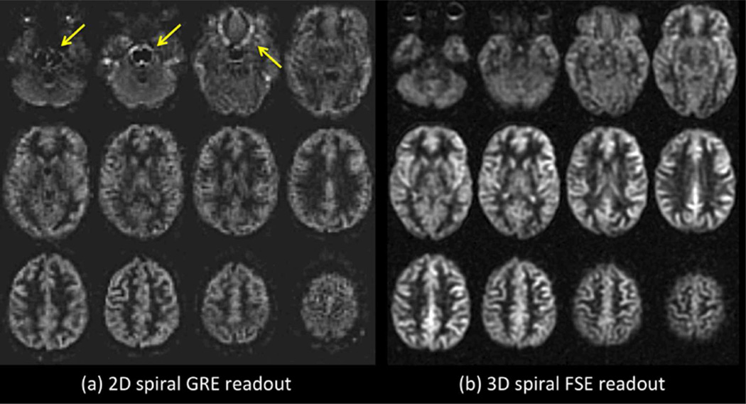
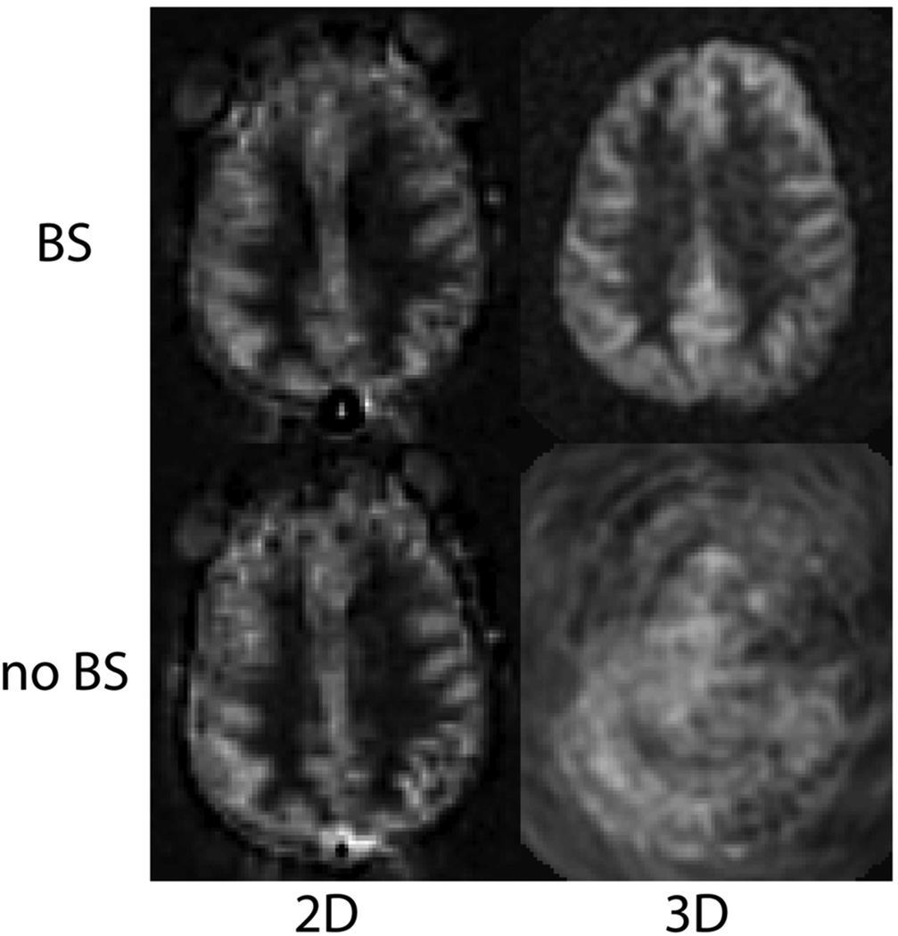
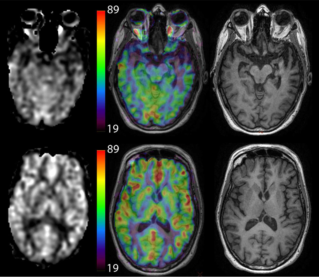
This review provides a summary statement of recommended implementations of arterial spin labeling (ASL) for clinical applications. It is a consensus of the ISMRM Perfusion Study Group and the European ASL in Dementia consortium, both of whom met to reach this consensus in October 2012 in Amsterdam. Although ASL continues to undergo rapid technical development, we believe that current ASL methods are robust and ready to provide useful clinical information, and that a consensus statement on recommended implementations will help the clinical community to adopt a standardized approach. In this review, we describe the major considerations and trade-offs in implementing an ASL protocol and provide specific recommendations for a standard approach. Our conclusion is that as an optimal default implementation, we recommend pseudo-continuous labeling, background suppression, a segmented three-dimensional readout without vascular crushing gradients, and calculation and presentation of both label/control difference images and cerebral blood flow in absolute units using a simplified model.

Keywords: arterial spin labeling; cerebral blood flow; perfusion.


Figures

Figure 1
Figure 1
Example of whole brain ASL imaging of cerebral blood flow at 3T using the recommended parameters in a normal subject, highlighting the typical image quality and expected contrast between gray and white matter. 101×84mm (300 × 300 DPI)
Figure 2
Figure 2
Schematic diagram of imaging and labeling regions for CASL/PCASL and PASL. In CASL/PCASL, labeling occurs as blood flow through a single labeling plane, while in PASL, a slab of tissue, including arterial blood, is labeled. 297×420mm (300 × 300 DPI)
Figure 3
Figure 3
(a) Example of poor PCASL labeling within the right anterior circulation due to poor labeling of the right internal carotid artery (ICA). Note the loss of ASL signal confined to this territory without compensatory collateral flow. In this case, confirmation was obtained with a (b) normal dynamic susceptibility contrast CBF map and (c) normal MR angiogram of the circle of Willis. (d) CT angiogram demonstrates surgical clips in the region of the right ICA (arrows), which may have been responsible for the poor labeling due to susceptibility effects. 60×35mm (300 × 300 DPI)
Figure 4
Figure 4
Timing diagram for CASL/PCASL and PASL. For QUIPSS II PASL, TI1 is the bolus duration, and is analogous to the labeling duration in CASL/PCASL. The post labeling delay (PLD) in CASL/PCASL is analogous to the quantity (TI-TI1) in QUIPSS II PASL. 297×420mm (300 × 300 DPI)
Figure 5
Figure 5
(a) 2D versus (b) 3D readout ASL imaging in a normal subject. Both images were acquired with approximately 5 min of imaging at 3T with PCASL labeling (label duration of 1.5 sec and a post-label delay of 2 sec). The 2D readout method was a single-shot gradient echo spiral. The 3D readout was a segmented stack-of-spirals FSE. Note the artifacts associated with the 2D single shot method in regions of high susceptibility (arrows). Parallel imaging approaches could be used to improve such artifacts associated with single-shot gradient echo imaging. 68×36mm (300 × 300 DPI)
Figure 6
Figure 6
PCASL images acquired using 2D single shot, and 3D segmented spiral readouts, with and without background suppression. 80×84mm (300 × 300 DPI)
Figure 7
Figure 7
Intraluminal ASL signal within veins (yellow arrows) indicative of arteriovenous shunting in a patient with a dural AV fistula (black arrow). Use of vascular crushing may suppress such information, limiting the clinical value of ASL in this type of case. 152×106mm (300 × 300 DPI)
Figure 8
Figure 8
Example of different methods to display CBF information in a patient with semantic dementia (note the low CBF in the left temporal lobe). Images on the left are CBF maps, while the center shows CBF maps using a color map overlaid on high-resolution T1-weighted images, which are show separately on the right. The color scale is in ml/min/100g. Color CBF maps may be displayed without anatomical underlay as well. 152×131mm (300 × 300 DPI)
Figure 9
Figure 9
Borderzone sign. These ASL subtraction images are from an 85 year-old man with dense left hemiparesis, acquired using PCASL with a labeling time of 1500 ms and a PLD of 1500 ms. Only the proximal portions of the arterial tree are present, indicating that the PLD was not long enough for the labeled spins to have reached the tissue, and that the ATT was prolonged bilaterally in this elderly patient. While longer PLD should improve the visualization of parenchymal CBF, it is not uncommon to see such a finding, known as the borderzone sign, in elderly patients with extremely delayed arrival times. 101×85mm (300 × 300 DPI)

Similar articles

Cited by

References

    1. Bokkers RP, Bremmer JP, van Berckel BN, Lammertsma AA, Hendrikse J, Pluim JP, Kappelle LJ, Boellaard R, Klijn CJ. Arterial spin labeling perfusion MRI at multiple delay times: a correlative study with H(2)(15)O positron emission tomography in patients with symptomatic carotid artery occlusion. Journal of cerebral blood flow and metabolism : official journal of the International Society of Cerebral Blood Flow and Metabolism. 2010;30(1):222–229. - PMC - PubMed
    1. Xu G, Rowley HA, Wu G, Alsop DC, Shankaranarayanan A, Dowling M, Christian BT, Oakes TR, Johnson SC. Reliability and precision of pseudo-continuous arterial spin labeling perfusion MRI on 3.0 T and comparison with 15O-water PET in elderly subjects at risk for Alzheimer's disease. NMR in biomedicine. 2010;23(3):286–293. - PMC - PubMed
    1. Gevers S, van Osch MJ, Bokkers RP, Kies DA, Teeuwisse WM, Majoie CB, Hendrikse J, Nederveen AJ. Intra- and multicenter reproducibility of pulsed, continuous and pseudo-continuous arterial spin labeling methods for measuring cerebral perfusion. Journal of cerebral blood flow and metabolism : official journal of the International Society of Cerebral Blood Flow and Metabolism. 2011;31(8):1706–1715. - PMC - PubMed
    1. Petersen ET, Mouridsen K, Golay X. The QUASAR reproducibility study, Part II: Results from a multi-center Arterial Spin Labeling test-retest study. NeuroImage. 2010;49(1):104–113. - PMC - PubMed
    1. Detre JA, Rao H, Wang DJ, Chen YF, Wang Z. Applications of arterial spin labeled MRI in the brain. Journal of magnetic resonance imaging : JMRI. 2012;35(5):1026–1037. - PMC - PubMed

Publication types