. 2014 Apr;141(8):1715-25.
doi: 10.1242/dev.095430.

Development of five digits is controlled by a bipartite long-range cis-regulator

Affiliations

Development of five digits is controlled by a bipartite long-range cis-regulator

Laura A Lettice et al. Development. 2014 Apr.

Abstract

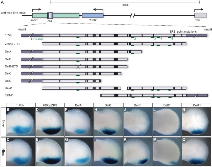
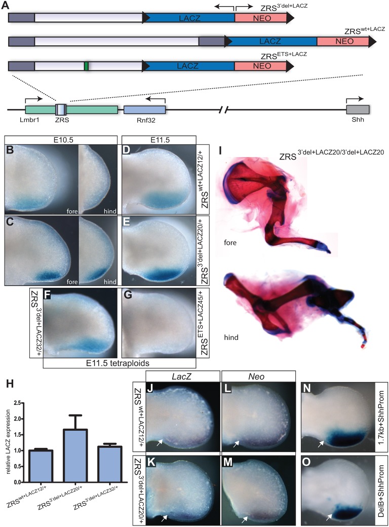
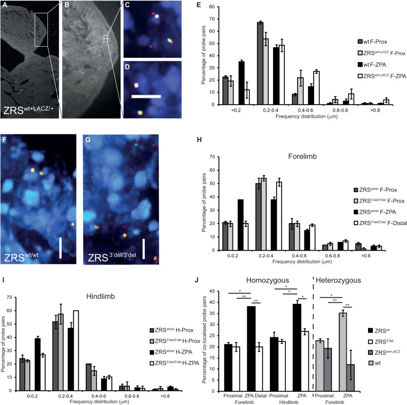
Conservation within intergenic DNA often highlights regulatory elements that control gene expression from a long range. How conservation within a single element relates to regulatory information and how internal composition relates to function is unknown. Here, we examine the structural features of the highly conserved ZRS (also called MFCS1) cis-regulator responsible for the spatiotemporal control of Shh in the limb bud. By systematically dissecting the ZRS, both in transgenic assays and within in the endogenous locus, we show that the ZRS is, in effect, composed of two distinct domains of activity: one domain directs spatiotemporal activity but functions predominantly from a short range, whereas a second domain is required to promote long-range activity. We show further that these two domains encode activities that are highly integrated and that the second domain is crucial in promoting the chromosomal conformational changes correlated with gene activity. During limb bud development, these activities encoded by the ZRS are interpreted differently by the fore limbs and the hind limbs; in the absence of the second domain there is no Shh activity in the fore limb, and in the hind limb low levels of Shh lead to a variant digit pattern ranging from two to four digits. Hence, in the embryo, the second domain stabilises the developmental programme providing a buffer for SHH morphogen activity and this ensures that five digits form in both sets of limbs.

Keywords: Limb development; Long-range regulation; Mouse; Sonic hedgehog; ZRS.


Figures

Fig. 1.
Fig. 1.
Deletion analysis of the ZRS cis-regulator. (A) Diagram of the Shh locus including the upstream gene desert and the position of the ZRS within an intron of the Lmbr1 gene. An expanded view of the 1.7-kb HindIII genomic fragment containing the 780-bp highly conserved ZRS is shown with the position of the point mutations (bars) and the ETS binding sites (green ovals) marked. The deletion constructs used in the transgenic analysis are shown below. (B-O) Expression of the lacZ gene in E11.5 embryonic limb buds (fore limbs in the top row and hind limbs in the bottom panel) for the 1.7-kb wild-type fragment (B,C), the conserved 780-bp ZRS (D,E), the DelA deletion (F,G), the DelB deletion (H,I), the DelC deletion (J,K), the DelD deletion (L,M) and the Del41 deletion (N,O). No expression was observed for the DelB-ETS or the 3′ END construct.
Fig. 2.
Fig. 2.
Mutational analysis within the ZRS. (A) The modified ZRS constructs used in transgenic analyses. The positions of the point mutations are marked by bars and M100081 and Hx highlighted in green and red, respectively. The sequence changes made within the Hx domain are also shown in red. (B-Q) Expression of the lacZ gene in E11.5 embryonic limb buds (fore limbs in the top row of each and hind limbs below) for the construct carrying the Hx mutation (B,C) (data from Lettice et al., 2008), the Flip80 construct (D,E), the Flip80+Hx construct (F,G), the Flip80+REP3A construct (H,I), the Flip49 construct (J,K), the Flip49+REP3A (L,M), the Core fragment (N,O) and the Core fragment carrying the M100081 mutation (P,Q). No expression was observed for the 3′END+Hx, the REP3A, REP3B, REP5 or the M100081+REP3A constructs.
Fig. 3.
Fig. 3.
Analysis of the targeted ZRS locus. (A) The three targeting constructs used to replace the endogenous ZRS. Each contains the lacZ gene (in the opposite orientation to the direction of Lmbr1 transcription) and the neoR (NEO) gene surrounded by LoxP sites (black triangles). The ZRSwt+LACZ targeted allele contains the 1.7-kb wild-type ZRS fragment. The ZRS3′del+LACZ allele contains a deletion of the 3′ end, retaining the DelB fragment and the ZRSETS+LACZ allele carries the additional mutation at the ETS1/GABPα binding site (represented by the green rectangle). (B-E) Limb expression of the lacZ reporter gene in the following mouse lines: the wild-type line ZRSwt+LACZ12 (B,D) and the 3′ deletion line ZRS3′del+LACZ20 (C,E). (B,C) ∼E10.5 limb buds, with fore limbs in the left panel and hind limbs in the right panel. (D,E) E11.5 hind limbs. (F,G) The analysis in tetraploid complementation embryos of lacZ expression in limb buds from the ZRS3′del+LACZ32 (F) and in ZRSETS+LACZ45 limb buds (G). (H) Graph showing the analysis by qRT-PCR of the levels of lacZ RNA present in the limb buds of ZRSwt+LACZ12 and the two 3′ deletion lines ZRS3′del+LACZ20 and ZRS3′del l+LACZ32. Mean±s.e.m. (I) Mutant limbs from an E17.5 embryo homozygous for ZRS3′del+LACZ20/3′del+LACZ20 showing the severe distal truncations in the fore limbs (upper) and hind limbs (lower). (J-O) The effect of different promoters on expression. (J,K) In situ expression of lacZ (driven by a β-globin minimal promoter). (L,M) Expression of NeoR (driven by the PGK promoter). (J,L) Limbs from the wild-type line ZRSwt+LACZ12. (K,M) Limbs from ZRS3′del l+LACZ20. (N,O) Limbs from transgenic embryos carrying lacZ driven by the Shh promoter and either 1.7-kb ZRS (N) or the truncated DelB (O). The proximal extent of expression is marked in all cases by a white arrow.
Fig. 4.
Fig. 4.
Analysis of limb development in the ZRS deletion mutant. (A) Mutations made in the ZRS after removing the lacZ reporter and the NeomycinR (NEO) selectable marker genes with Cre recombinase leaving behind a single LoxP site (black triangle). (B-V) All of the images shown are taken from ZRS3′del20 derived from the ZRS3′del+LACZ20 line. (B-D) The long bones of the hind limbs are shown in ZRSwt/wt (B) and ZRS3′del/3′del (C,D). (E-F‴) The range of digit number that forms in the hind limb in the ZRS3′del/3′del mutants (F-F‴) compared with the wild-type pattern produced by ZRSwt/wt (E). (G-N) Expression analysis in E11.5 embryos of Shh (G-J) and Ptc (K-N) in wild-type (wt) mice and ZRS3′del/3′del fore and hind limbs as indicated. For Shh expression, n=15 wt and 10 mutants and for Ptc expression n=16 wt and 6 mutants. Note the low expression of Shh but in a normal pattern (indicated by the dotted line) (J) and the lower levels of Ptc (N) compared with wt. (O-V) At an earlier stage, E10.5, expression of Shh (O,P) and Ptc (S,T) is detected in wt limbs. However, expression of both Shh (Q,R) and Ptc (U,V) is undetectable in ZRS3′del/3′del limbs. For Shh expression, n=14 wt and 12 mutants and for Ptc expression n=9 wt and 5 mutants. (W) Levels of Shh expressed in the individual limb bud pairs of wild-type, heterozygous (Shhnull/+) and mutant (ZRS3′del/3′del) fore and hind limb buds analysed by qRT-PCR. The mean±s.e.m. is plotted. These values were subjected to a non parametric Mann–Whitney U-test (**P<0.01).
Fig. 5.
Fig. 5.
Analysis of chromosome conformation by FISH. (A,B) Low magnification image of a DAPI-stained section from a ZRSwt+LACZ/+ embryo (A) at E11.5 and a close up of the posterior limb bud, with the ZPA highlighted by staining for the SHH protein (B). (C,D) Four-colour FISH images showing two probe pairs from within the ZPA stained for Shh (red), the ZRS (green) and the lacZ reporter (white). The lacZ staining highlights the ZRSwt+LACZ allele. (E) Graph of the distribution of interprobe distances between Shh and ZRS in the wild-type allele and the ZRSwt+LACZ allele in the proximal limb bud and in the ZPA of a ZRSwt+LACZ/+ embryo. (F,G) Three-colour FISH images showing probe pairs in the ZPA from ZRSwt/wt (F) and ZRS3′del/3′del (G) embryos (Shh stained red and ZRS with green). (H,I) Distribution of interprobe distances between Shh and the ZRS in the proximal and distal fore limb (H) and hind limb (I) buds of ZRSwt/wt and ZRS3′del/3′del embryos. (J) Summary of the measurements and comparison of colocalised probe pairs in ZRSwt/wt, ZRS3′del/3′del and ZRSwt+LACZ/+. Significant comparisons are indicated (*P<0.05, **P<0.01 compared with the relevant wild-type ZPA sample). Significantly greater Shh-ZRS colocalisation is identified in the ZPA of fore limb and hind limb in ZRSwt compared with ZRS3′del, ZRSwt+LACZ and proximal wild type. Loss of colocalisation at the lacZ allele in distal posterior nuclei corresponds to an increased proportion of probe pair distances greater than 400 nm. No detectable difference in probe colocalisation occurs in the proximal nuclei. Between 100 and 175 loci were measured for each tissue and probe pair. Tables of the results of the statistical tests are shown in supplementary material Tables S3-S6. F-Prox, fore limb proximal; F-ZPA, fore limb ZPA; F-Distal, fore limb distal posterior region; H-prox, hind limb proximal; H-ZPA, hind limb ZPA; Wt, wild type. Scale bars: 5 µm.

References

    1. Ahn S., Joyner A. L. (2004). Dynamic changes in the response of cells to positive hedgehog signalling during mouse limb patterning. Cell 118, 505-516 10.1016/j.cell.2004.07.023 - DOI - PubMed
    1. Amano T., Sagai T., Tanabe H., Mizushina Y., Nakazawa H., Shiroishi T. (2009). Chromosomal dynamics at the Shh locus: limb bud-specific differential regulation of competence and active transcription. Dev. Cell 16, 47-57 10.1016/j.devcel.2008.11.011 - DOI - PubMed
    1. Anderson E., Peluso S., Lettice L. A., Hill R. E. (2012). Human limb abnormalities caused by disruption of hedgehog signaling. Trends Genet. 28, 364-373 10.1016/j.tig.2012.03.012 - DOI - PubMed
    1. Araki K., Araki M., Miyazaki J., Vassalli P. (1995). Site-specific recombination of a transgene in fertilized eggs by transient expression of Cre recombinase. Proc. Natl Acad. Sci. U.S.A. 92, 160-164 10.1073/pnas.92.1.160 - DOI - PMC - PubMed
    1. Blanc I., Bach A., Robert B. (2002). Unusual pattern of Sonic hedgehog expression in the polydactylous mouse mutant Hemimelic extra-toes. Int. J. Dev. Biol. 46, 969-974 - PubMed

Publication types

MeSH terms