

Evolving character of chronic central nervous system HIV infection
- PMID: 24715483
- PMCID: PMC4120280
- DOI: 10.1055/s-0034-1372337
Evolving character of chronic central nervous system HIV infection
Abstract
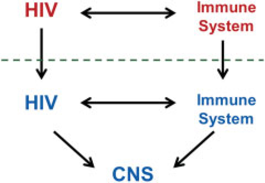
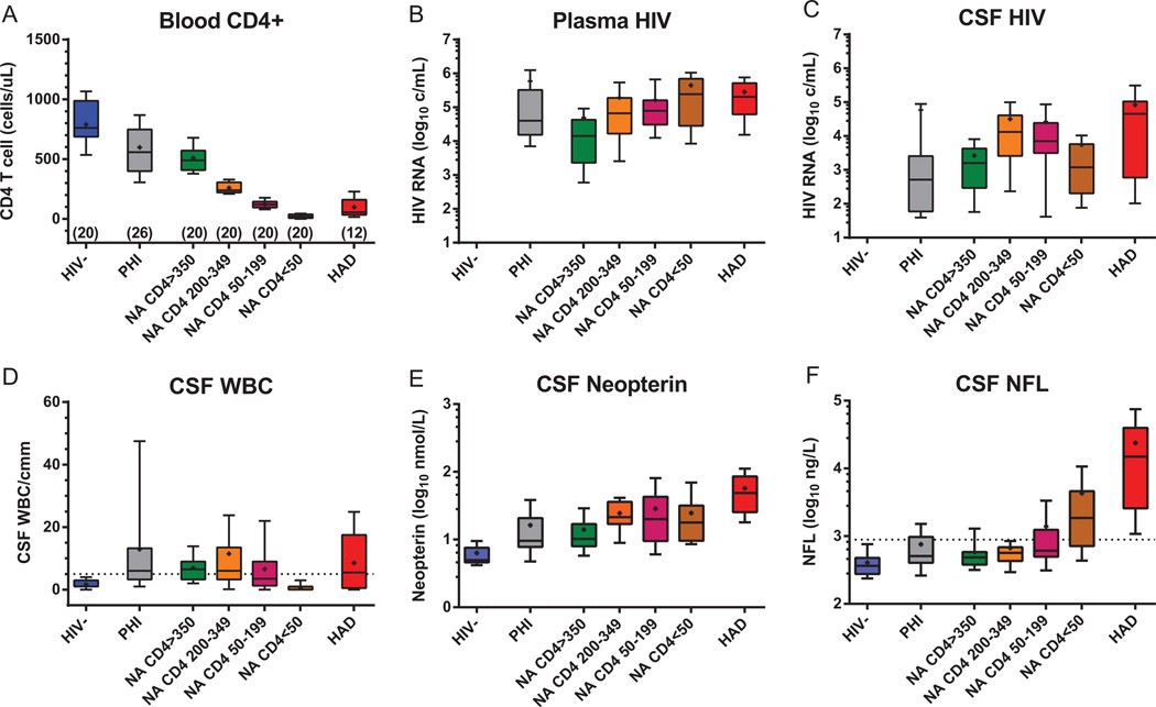
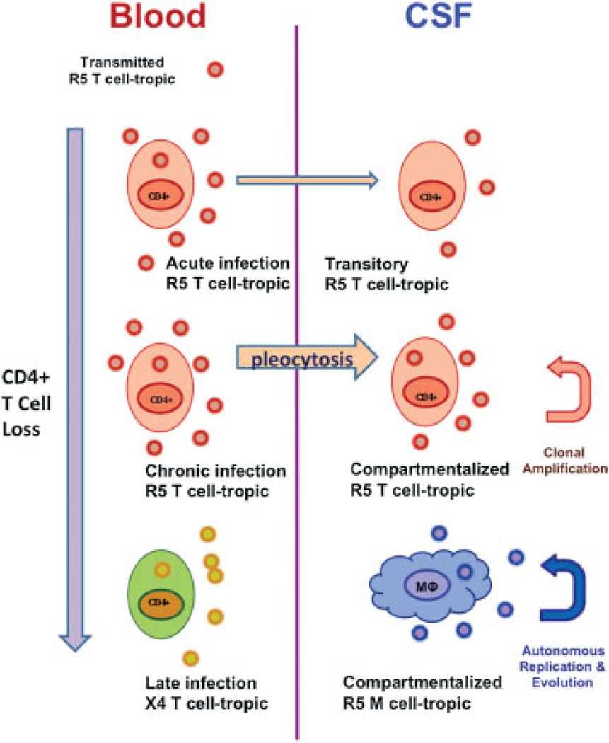
Human immunodeficiency virus type 1 (HIV-1) infection of the central nervous system (CNS) begins early in systemic infection and continues throughout its untreated course. Despite a common cerebrospinal fluid inflammatory response, it is usually neurologically asymptomatic for much of this course, but can evolve in some individuals to HIV-associated dementia (HAD), a severe encephalopathy with characteristic cognitive and motor dysfunction. While widespread use of combination antiretroviral therapy (ART) has led to a marked decline in both the CNS infection and its neurologic severe consequence, HAD continues to afflict individuals presenting with advanced systemic infection in the developed world and a larger number in resource-poor settings where ART is more restricted. Additionally, milder CNS injury and dysfunction have broader prevalence, including in those treated with ART. Here we review the history and evolving nomenclature of HAD, its viral pathogenesis, clinical presentation and diagnosis, and treatment.
Thieme Medical Publishers 333 Seventh Avenue, New York, NY 10001, USA.
Conflict of interest statement
RWP is a consultant to Merck and Co and has received an honorarium and travel support from Abbott Laboratories for meeting presentation. MG has received research grants from Abbott, Baxter, Bristol-Myers Squibb, Gilead Sciences, GlaxoSmithKline, Merck, Pfizer, Roche, and Tibotec. He has received honoraria as speaker and/or advisor from Abbott/Abbvie, Bioinvent, Boehringer-Ingelheim, Bristol-Myers Squibb, Gilead Sciences, GlaxoSmithKline, Janssen-Cilag, Merck, Pfizer, Roche, and Tibotec/Janssen. SS has received an honorarium and travel support from AbbVie, Inc.
Figures
Similar articles
-
Global developments in HIV neurology.Handb Clin Neurol. 2018;152:265-287. doi: 10.1016/B978-0-444-63849-6.00019-0. Handb Clin Neurol. 2018. PMID: 29604981 Review.
-
Antiretroviral therapy and central nervous system HIV type 1 infection.J Infect Dis. 2008 May 15;197 Suppl 3(Suppl 3):S294-306. doi: 10.1086/533419. J Infect Dis. 2008. PMID: 18447615 Free PMC article. Review.
-
Opportunistic Neurologic Infections in Patients with Acquired Immunodeficiency Syndrome (AIDS).Curr Neurol Neurosci Rep. 2016 Jan;16(1):10. doi: 10.1007/s11910-015-0603-8. Curr Neurol Neurosci Rep. 2016. PMID: 26747443 Review.
-
HIV-associated opportunistic infections of the CNS.Lancet Neurol. 2012 Jul;11(7):605-17. doi: 10.1016/S1474-4422(12)70098-4. Lancet Neurol. 2012. PMID: 22710754 Review.
-
Central Nervous System Infections in the Immunocompromised Adult Presenting to the Emergency Department.Emerg Med Clin North Am. 2021 Feb;39(1):101-121. doi: 10.1016/j.emc.2020.09.006. Epub 2020 Nov 5. Emerg Med Clin North Am. 2021. PMID: 33218652 Review.
Cited by
-
No Association Between Human Immunodeficiency Virus Infections And Dementia: A Nationwide Cohort Study In Taiwan.Neuropsychiatr Dis Treat. 2019 Nov 12;15:3155-3166. doi: 10.2147/NDT.S225584. eCollection 2019. Neuropsychiatr Dis Treat. 2019. PMID: 31814723 Free PMC article.
-
Changes in cerebrospinal fluid proteins across the spectrum of untreated and treated chronic HIV-1 infection.PLoS Pathog. 2024 Sep 24;20(9):e1012470. doi: 10.1371/journal.ppat.1012470. eCollection 2024 Sep. PLoS Pathog. 2024. PMID: 39316609 Free PMC article.
-
Neuroinflammatory transcriptional programs induced in rhesus pre-frontal cortex white matter during acute SHIV infection.J Neuroinflammation. 2022 Oct 6;19(1):250. doi: 10.1186/s12974-022-02610-y. J Neuroinflammation. 2022. PMID: 36203187 Free PMC article.
-
Latent HIV-Exosomes Induce Mitochondrial Hyperfusion Due to Loss of Phosphorylated Dynamin-Related Protein 1 in Brain Endothelium.Mol Neurobiol. 2021 Jun;58(6):2974-2989. doi: 10.1007/s12035-021-02319-8. Epub 2021 Feb 14. Mol Neurobiol. 2021. PMID: 33586027 Free PMC article.
-
Developments in Neuroprotection for HIV-Associated Neurocognitive Disorders (HAND).Curr HIV/AIDS Rep. 2022 Oct;19(5):344-357. doi: 10.1007/s11904-022-00612-2. Epub 2022 Jul 22. Curr HIV/AIDS Rep. 2022. PMID: 35867211 Free PMC article. Review.
References
-
- Davis LE, Hjelle BL, Miller VE, et al. Early viral brain invasion in iatrogenic human immunodeficiency virus infection. Neurology. 1992;42(9):1736–1739. - PubMed
-
- Ellis RJ, Hsia K, Spector SA, et al. HIV Neurobehavioral Research Center Group. Cerebrospinal fluid human immunodeficiency virus type 1 RNA levels are elevated in neurocognitively impaired individuals with acquired immunodeficiency syndrome. Ann Neurol. 1997;42(5):679–688. - PubMed
Publication types
MeSH terms
Substances
Grants and funding
LinkOut - more resources
Full Text Sources
Other Literature Sources
Medical
