. 2014 Apr 22;111(16):E1591-9.
doi: 10.1073/pnas.1321902111. Epub 2014 Apr 8.

The nature and extent of contributions by defective ribosome products to the HLA peptidome

Affiliations

The nature and extent of contributions by defective ribosome products to the HLA peptidome

Dmitry Bourdetsky et al. Proc Natl Acad Sci U S A. .

Abstract

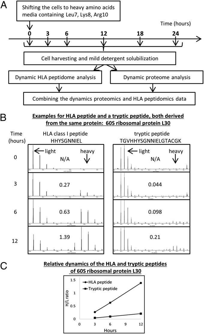
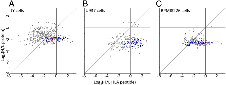
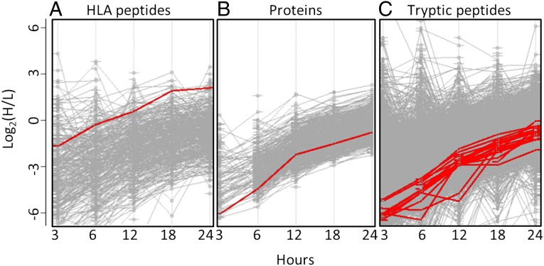
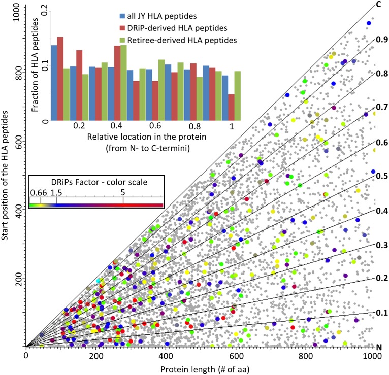
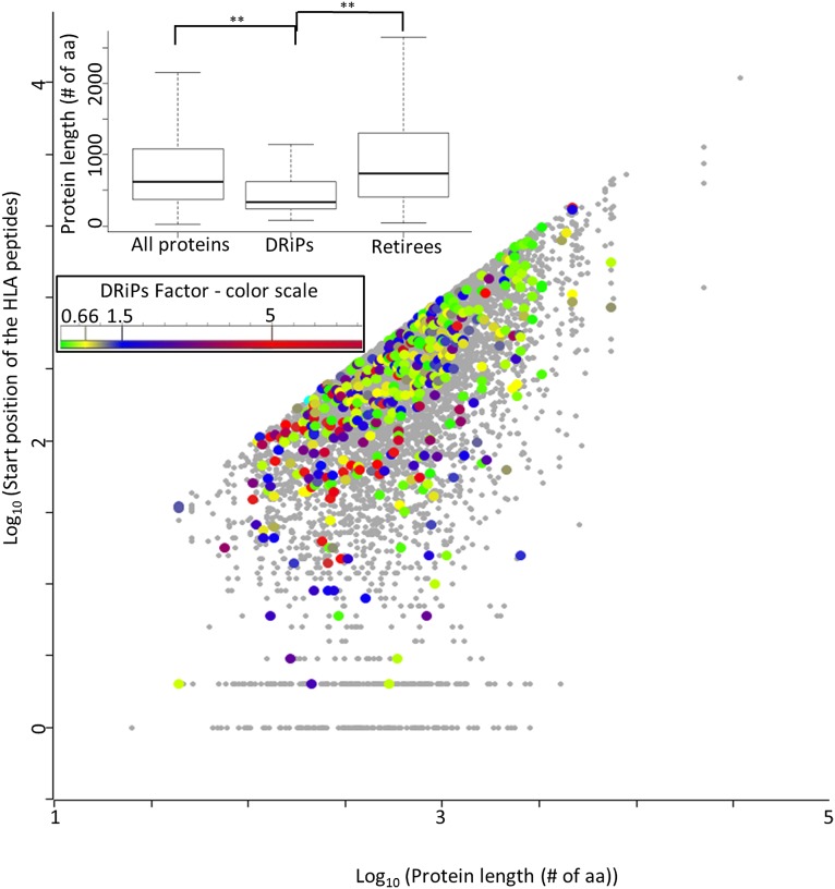
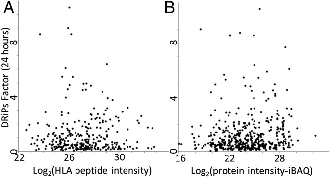
MHC class I peptides are products of endogenous cellular protein degradation. Their prompt presentation, after rapid degradation of their newly synthesized source proteins, is needed to alert the immune system during pathogen infection. A possible source for such rapidly degrading proteins can be defective ribosome products (DRiPs), which include polypeptides produced as part of the pioneer round of translation, premature translation termination, and proteins failing to fold properly or to assemble into their multisubunit protein complexes. However, the identities and relative contribution to the MHC peptidome of these mature or newly synthesized and rapidly degraded cellular proteins is not well understood. To clarify these issues, we used dynamic stable isotope labeling by amino acids in cell culture to define the relative rates of synthesis of the HLA class I peptidomes and the source proteomes of three cultured human hematopoietic cell lines. Large numbers of HLA class I peptides were observed to be derived from DRiPs, defined here as HLA peptides that shift from their light to heavy isotope forms faster than their source proteins. Specific groups of proteins, such as ribosomal and T-complex protein 1 (TCP-1), contributed a disproportionately large number of DRiPs to the HLA peptidomes. Furthermore, no significant preference was observed for HLA peptides derived from the amino terminal regions of the proteins, suggesting that the contribution of products of premature translation termination was minimal. Thus, the most likely sources of DRiPs-derived HLA peptides are full-sized, misassembled, and surplus subunits of large protein complexes.

Keywords: DRiPome; dynamic SILAC; immunopeptidome.


Conflict of interest statement

The authors declare no conflict of interest.

Figures

Fig. 1.
Fig. 1.
Experimental flowchart depicting the isolation and dynamic SILAC analysis of the proteomes (through its tryptic peptides) and the HLA class I peptidomes of the same cultured cells. The proteins and HLA peptides dynamics were defined by shifting the growth media from light to heavy amino acids (A). Multiple time-point mass spectra of an HLA and a tryptic peptide, both derived from the same protein, illustrating the shift from light to heavy form (calculated H/L ratios are presented in the boxes) (B). Graphical representation of H/L ratios of the same HLA and the tryptic peptides from the 60S ribosomal L30 protein (C).
Fig. 2.
Fig. 2.
Relative H/L ratios of HLA peptides and of their source proteins at the 12-h time points of the JY (A), U937 (B), and RPMI8226 (C) cells. The different subunits of ribosomes are labeled as blue circles and the subunits of TCP-1 as red squares.
Fig. 3.
Fig. 3.
Comparative dynamic SILAC data of the proteome and the HLA peptidome of the JY cells: kinetics of HLA peptides (created by Perseus software) (A); protein kinetics, as median of their tryptic peptides kinetics (B); and kinetics of individual tryptic peptides (C). Each gray line indicates the kinetics of one HLA peptide (A), one protein (B) or one tryptic peptide (C). As an example, the bold lines represent the kinetics of an HLA peptide derived from the T-complex protein 1 subunit eta and of the tryptic peptides of the same protein.
Fig. 4.
Fig. 4.
Relative locations of HLA peptides within the first 1,000 amino acids of their source proteins in the JY cells. The small gray dots represent HLA peptides and their source proteins for which the DRiPs factor was not defined. The colored dots indicate HLA peptides with defined DRiPs factors, with the color indicating the DRiPs factor scale (Inset). Diagonal lines represent the percentile values of the locations of the HLA peptides within their source proteins. The Inset bar graph displays the fractions of high DRiPs factor peptides (derived from the certain DRiPs), low DRiPs factor peptides (derived from the certain retirees), and the entire list of HLA peptides, according to their relative locations within their source proteins.
Fig. 5.
Fig. 5.
The position of HLA peptides within the entire lengths of their source proteins, in the JY cells. The colors indicate the DRiPs factors of the HLA peptides according to the DRiPs factor scale (Inset). The statistical significance between the protein's length distribution of all proteins, DRiPs, and retiree, were evaluated by the Mann–Whitney test (**P < 0.001).
Fig. 6.
Fig. 6.
Correlation between the DRiPs factors and the cellular expression levels of the HLA peptides (A) and of the proteins (B). The cellular levels of the proteins were defined as their iBAQ intensities.

References

    1. Esquivel F, Yewdell J, Bennink J. RMA/S cells present endogenously synthesized cytosolic proteins to class I-restricted cytotoxic T lymphocytes. J Exp Med. 1992;175(1):163–168. - PMC - PubMed
    1. Kim Y, Yewdell JW, Sette A, Peters B. Positional bias of MHC class I restricted T-cell epitopes in viral antigens is likely due to a bias in conservation. PLOS Comput Biol. 2013;9(1):e1002884. - PMC - PubMed
    1. Farfán-Arribas DJ, Stern LJ, Rock KL. Using intein catalysis to probe the origin of major histocompatibility complex class I-presented peptides. Proc Natl Acad Sci USA. 2012;109(42):16998–17003. - PMC - PubMed
    1. Yewdell JW, Antón LC, Bennink JR. Defective ribosomal products (DRiPs): A major source of antigenic peptides for MHC class I molecules? J Immunol. 1996;157(5):1823–1826. - PubMed
    1. Reits EA, Vos JC, Grommé M, Neefjes J. The major substrates for TAP in vivo are derived from newly synthesized proteins. Nature. 2000;404(6779):774–778. - PubMed

Publication types