. 2014 Apr 22;111(16):5878-83.
doi: 10.1073/pnas.1322473111. Epub 2014 Apr 8.

Outer membrane β-barrel protein folding is physically controlled by periplasmic lipid head groups and BamA

Affiliations

Outer membrane β-barrel protein folding is physically controlled by periplasmic lipid head groups and BamA

Dennis Gessmann et al. Proc Natl Acad Sci U S A. .

Abstract

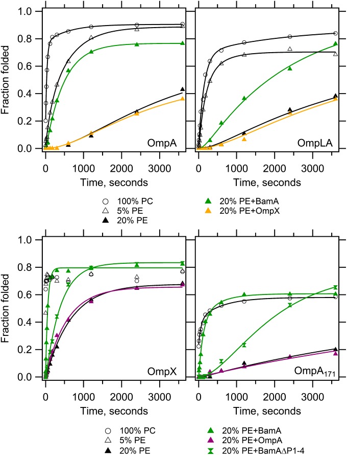
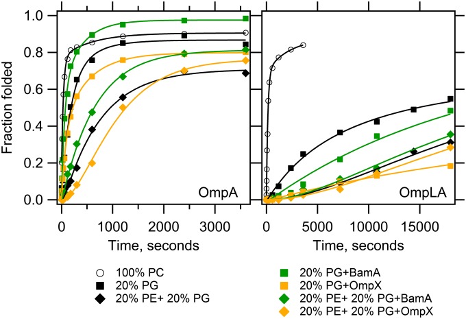
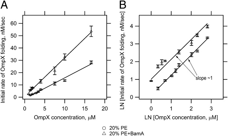
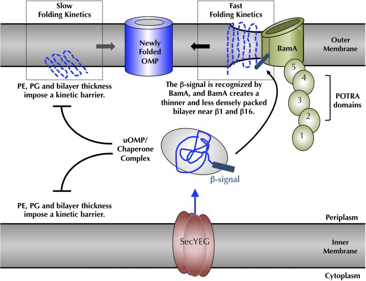
Outer membrane β-barrel proteins (OMPs) are crucial for numerous cellular processes in prokaryotes and eukaryotes. Despite extensive studies on OMP biogenesis, it is unclear why OMPs require assembly machineries to fold into their native outer membranes, as they are capable of folding quickly and efficiently through an intrinsic folding pathway in vitro. By investigating the folding of several bacterial OMPs using membranes with naturally occurring Escherichia coli lipids, we show that phosphoethanolamine and phosphoglycerol head groups impose a kinetic barrier to OMP folding. The kinetic retardation of OMP folding places a strong negative pressure against spontaneous incorporation of OMPs into inner bacterial membranes, which would dissipate the proton motive force and undoubtedly kill bacteria. We further show that prefolded β-barrel assembly machinery subunit A (BamA), the evolutionarily conserved, central subunit of the BAM complex, accelerates OMP folding by lowering the kinetic barrier imposed by phosphoethanolamine head groups. Our results suggest that OMP assembly machineries are required in vivo to enable physical control over the spontaneously occurring OMP folding reaction in the periplasm. Mechanistic studies further allowed us to derive a model for BamA function, which explains how OMP assembly can be conserved between prokaryotes and eukaryotes.

Keywords: beta-barrel transmembrane protein; membrane protein folding.


Conflict of interest statement

The authors declare no conflict of interest.

Figures

Fig. 1.
Fig. 1.
BamA lowers the kinetic barrier to OMP folding imposed by PE head groups. Unfolded OMPs were folded into LUVs containing 100% PC or 95% PC + 5% PE or 80% PC + 20% PE in the absence or presence of one of the following prefolded OMPs as indicated: BamA, BamAΔP1-4, OmpA or OmpX. The client OMP is indicated in each panel, and its total concentration was 4 or 2 μM without or with prefolded OMP, respectively, at a constant lipid:protein mole ratio of 800:1. Samples were taken at indicated times and quenched with SDS/PAGE loading buffer. SDS/PAGE gels (SI Appendix, Fig. S2) were analyzed by densitometry and fraction folded was plotted as the ratio of the folded band intensity divided by the boiled band intensity. Lines indicate fitted curves; black, no prefolded OMP; green, prefolded BamA or BamAΔP1-4; orange, prefolded OmpX; magenta, prefolded OmpA. Each experiment was conducted in triplicate, and one representative data set is shown for each condition. SI Appendix, Fig. S3 shows representative data that includes the extended time points collected for the slower kinetic data, demonstrating that folding was collected to saturation. The average fit values with SDs are reported in SI Appendix, Tables S1–S4 and Fig. S4.
Fig. 2.
Fig. 2.
BamA functionality is modulated by PE and PG head groups. Unfolded OmpA and OmpLA were folded into PC LUVs composed of 20% PG or 20% PE + 20% PG with or without prefolded BamA or OmpX. The client OMP is indicated in each panel, and its total concentration was 4 or 2 μM without or with prefolded OMP, respectively, at a constant lipid:protein ratio of 800:1. Samples were recorded, analyzed and fitted as described in Fig. 1. Kinetic traces of OMP folding into 100% PC LUVs containing no prefolded OMP are replotted from Fig. 1 for comparison. Lines indicate fitted curves; black, no prefolded OMP; green, prefolded BamA; orange, prefolded OmpX. Each experiment was conducted in triplicate, and one representative data set is shown for each condition. Average fit values with SDs are reported in SI Appendix, Tables S1 and S2 and Fig. S4. Longer time points for the slow OmpLA transients are shown in SI Appendix, Fig. S5.
Fig. 3.
Fig. 3.
BamA catalyzes the folding reaction of OmpX. (A) Initial rates of OmpX folding as a function of OmpX concentration. (B) Double logarithmic plot of initial OmpX folding rates as a function of OmpX concentration. OmpX was folded into PC LUVs containing 20% PE with or without prefolded BamA at different final OmpX concentrations. The lipid concentration was 1.6 mM, and the final concentration of prefolded BamA was ∼1.6 μM. SI Appendix, Fig. S6 shows representative kinetic traces of OmpX folding with and without prefolded BamA that were used to determine the initial OmpX folding rates. Experiments were conducted in triplicate and samples were recorded, analyzed and fitted as described in Fig. 1.
Fig. 4.
Fig. 4.
The C-terminal phenylalanine of the OMP β-signal is required for folding via BamA. Unfolded OmpA171ΔPhe and OmpLAΔPhe were folded into PC LUVs with or without prefolded BamA. The total concentration of the target OMP was 4 or 2 μM without or with prefolded BamA, respectively, at a constant lipid:protein ratio of 800:1. Data were recorded, analyzed, and fitted as described in Fig. 1. Kinetic traces for the corresponding wild-type OMP are replotted from Fig, 1. Fit values are reported in SI Appendix, Tables S2 and S4 and Fig. S7. Lines indicate fitted curves; black, no prefolded OMP; green, prefolded BamA. Longer time points for the slow transients are shown in SI Appendix, Fig. S8.
Fig. 5.
Fig. 5.
Cellular consequences of the lipid-induced kinetic folding barrier and model for BamA-catalyzed folding. Folding of bacterial OMPs into the outer membrane of E. coli is kinetically prevented by the bilayer thickness and a densely packed membrane surface, caused by PE and PG head groups (, –39). At the outer membrane, BamA accelerates client OMP folding by the creation of local defects in and thinning of the membrane bilayer (32), which promote faster intrinsic folding kinetics (15). β-signals of client OMPs are recognized by BamA, which localizes clients to the site of the membrane defect. A high Km value of BamA for client OMPs together with favorable OMP folding free energies (49) drive the client OMPs to dissociate from BamA and move away into the membrane. This mechanistic view indicates how bacterial and mitochondrial OMPs can be recognized and folded by the eukaryotic BamA homologs (44, 45). POTRA, polypeptide-transport-associated.

References

    1. Tamm LK, Hong H, Liang B. Folding and assembly of beta-barrel membrane proteins. Biochim Biophys Acta. 2004;1666(1-2):250–263. - PubMed
    1. Wimley WC. The versatile beta-barrel membrane protein. Curr Opin Struct Biol. 2003;13(4):404–411. - PubMed
    1. Nikaido H. Outer membrane barrier as a mechanism of antimicrobial resistance. Antimicrob Agents Chemother. 1989;33(11):1831–1836. - PMC - PubMed
    1. Bajaj H, et al. Antibiotic uptake through membrane channels: Role of Providencia stuartii OmpPst1 porin in carbapenem resistance. Biochemistry. 2012;51(51):10244–10249. - PubMed
    1. Manczak M, Reddy PH. Abnormal interaction of VDAC1 with amyloid beta and phosphorylated tau causes mitochondrial dysfunction in Alzheimer’s disease. Hum Mol Genet. 2012;21(23):5131–5146. - PMC - PubMed

Publication types

MeSH terms