

Showcase of Intraoperative 3D Imaging of the Sentinel Lymph Node in a Breast Cancer Patient using the New Freehand SPECT Technology
- PMID: 24715831
- PMCID: PMC3971815
- DOI: 10.1159/000345472
Showcase of Intraoperative 3D Imaging of the Sentinel Lymph Node in a Breast Cancer Patient using the New Freehand SPECT Technology
Abstract
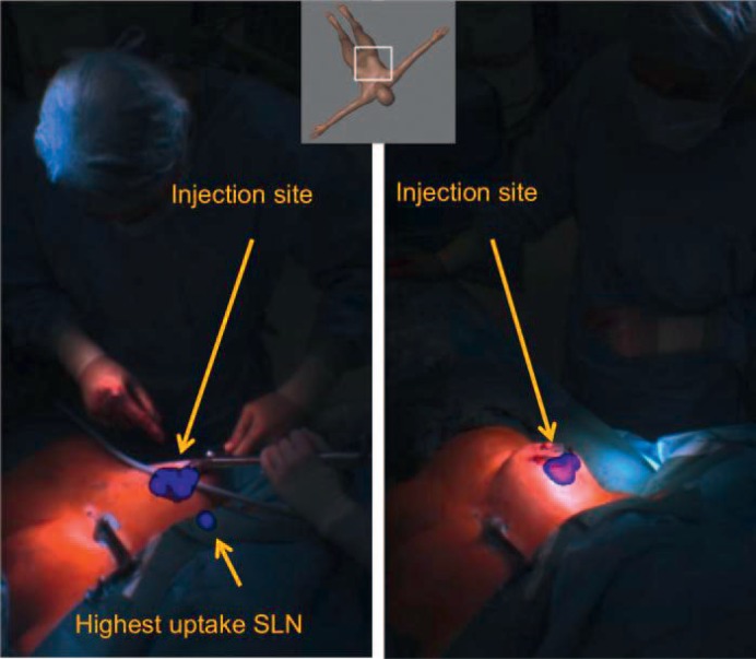
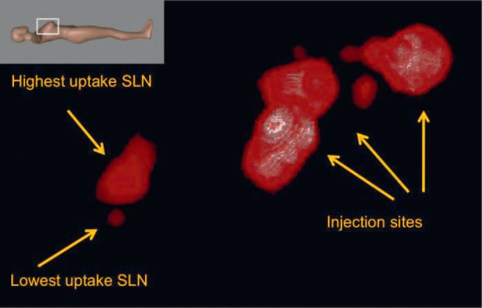
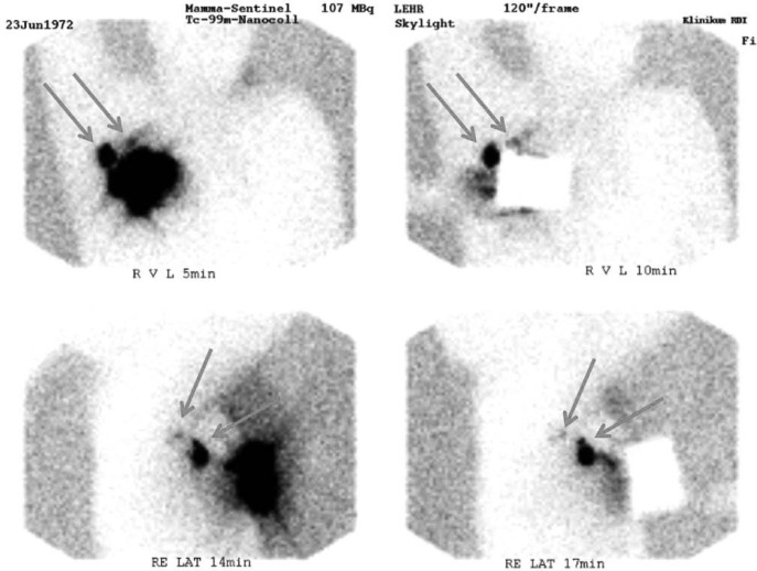
After the development of a hand-held intraoperative device for 3D real-time imaging of radioactively labeled sentinel lymph nodes in the human body, we present our first experience with the newest version of the freehand single-photon emission computed tomography (SPECT) technology in the operating room. The freehand SPECT system combines a gamma probe and an optical infrared positioning system, and provides surgeons with 3D imaging including exact depth information of the radioactive target. This technology was used intraoperatively in a female breast cancer patient to localize the axillary sentinel lymph nodes. The data obtained with freehand SPECT correlate well with conventional lymphoscintigraphy and with data collected using a conventional hand-held probe. By offering fast real-time intraoperative imaging, the new freehand SPECT system might facilitate the detection and removal of the sentinel lymph node(s) in certain situations and can be used for documentation and quality assurance purposes.
Dieser Bericht beschreibt die ersten klinischen Erfahrungen mit der neuesten Freehand-SPECT-Technologie, die zum intraoperativen Nachweis radioaktiv markierter Lymphknoten im menschlichen Körper entwickelt wurde. Die technische Grundlage basiert auf der Kombination einer konventionellen Gammasonde mit einem optischen Infrarot-Positionierungssystem. Der Operateur erhält eine räumliche Darstellung der Radioaktivität einschließlich der exakten Tiefenangabe im menschlichen Körper. Bei einer Mammakarzinompatientin wurde das System zum Auffinden des axillären Sentinellymphknotens verwendet. Die erhobenen Messwerte korrelieren sowohl mit den Ergebnissen einer konventionellen Lymphszintigraphie als auch mit den intraoperativen Messungen der mobilen Gammasonde. Die neue Freehand-SPECT-Technologie hat das Potential, dem Operateur durch die räumliche Echtzeitdarstellung der Radioaktivität das Auffinden und Entfernen des Sentinellymphknotens in bestimmten Situationen zu erleichtern und kann zu Zwecken der Dokumentation und Qualitätssicherung dienen.
Keywords: Breast cancer; Lymph node dissection; Quality assurance; Sentinel lymph node.
Figures
References
-
- Lyman GH, Giuliano AE, Somerfield MR, Benson AB, 3rd, Bodurka DC, Burstein HJ, Cochran AJ, Cody HS, 3rd, Edge SB, Galper S, Hayman JA, Kim TY, Perkins CL, Podoloff DA, Sivasubramaniam VH, Turner RR, Wahl R, Weaver DL, Wolff AC, Winer EP. American Society of Clinical Oncology guideline recommendations for sentinel lymph node biopsy in early-stage breast cancer. J Clin Oncol. 2005;23:7703–7720. - PubMed
-
- Schuman S, Walker G, Avisar E. Processing sentinel nodes: When and how many? Arch Surg. 2011;146:389–393. - PubMed
-
- Doting MH, Stiekema HM, de Vries J, Lemstra C, Hoekstra HJ, Vrieling M, Rietman L, Jager PL. Immediate dynamic lymphoscintigraphy delivers no additional value to lymphoscintigraphy 3 hr after tracer injection in sentinel lymph node biopsy in breast cancer patients. J Surg Oncol. 2007;95:469–475. - PubMed
-
- Sadeghi R, Forghani MN, Memar B, Abdollahi A, Zakavi SR, Mashhadi MT, Raziee HR, Tavassoli A, Kakhki VR. Comparison of pre-operative lymphoscintigraphy with inter-operative gamma probe and dye technique regarding the number of detected sentinel lymph nodes. Hell J Nucl Med. 2009;12:30–32. - PubMed
-
- Husarik DB, Steinert HC. Single-photon emission computed tomography/computed tomography for sentinel node mapping in breast cancer. Semin Nucl Med. 2007;37:29–33. - PubMed
LinkOut - more resources
Full Text Sources
