

Filariasis of the axilla in a patient returning from travel abroad: a case report
- PMID: 24715832
- PMCID: PMC3971795
- DOI: 10.1159/000345471
Filariasis of the axilla in a patient returning from travel abroad: a case report
Abstract
Background: The term filariasis comprises a group of parasitic infections caused by helminths belonging to different genera in the superfamily Filaroidea. The human parasites occur mainly in tropical and subtropical regions, but filariae are also found in temperate climates, where they can infect wild and domestic animals. Humans are rarely infected by these zoonotic parasites.
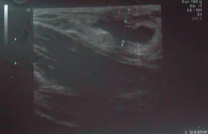
Patients and methods: A 55-year-old patient presented with a new-onset, subcutaneous, non-tender palpable mass in the right axilla. Ultrasonography showed a 1.3-cm, solid, singular encapsulated node. Sonography of the breast on both sides, axilla and lymphatic drainage on the left side, lymphatic drainage on the right side, and mammography on both sides were without pathological findings. The node was excised under local anesthesia as the patient refused minimal invasive biopsy.
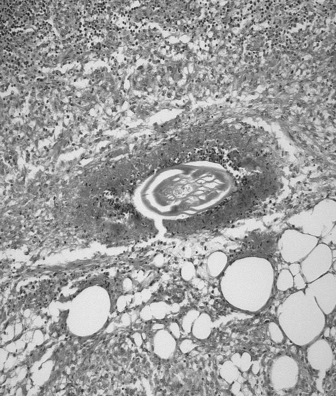
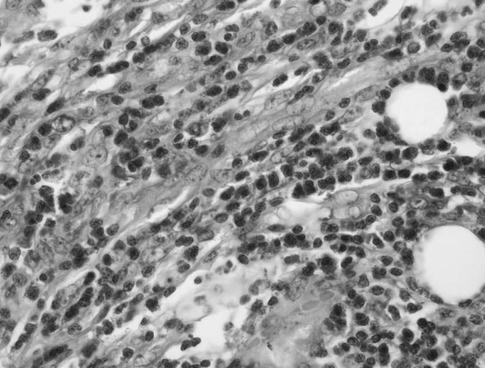
Results: On histopathological examination, the tail of a parasite of the group of filariae was found. The patient revealed that she had stayed in Africa and Malaysia for professional reasons. 6 months before the time of diagnosis, she had also suffered from a fever and poor general condition after a trip abroad. The patient was referred for further treatment to the Institute for Tropical Medicine at the University of Dusseldorf, where a treatment with ivermectin was conducted on the basis of positive staining with antibodies against filariae.
Conclusion: Our case demonstrates the importance of interdisciplinary collaboration between breast center, pathology, and other specialties such as microbiology and tropical medicine.
Hintergrund: Der Begriff Filariose umfasst eine Gruppe von parasitären Infektionen, die von Helminthen verschiedener Gattungen der Überfamilie der Filaroidea verursacht werden. Die Humanparasiten kommen hauptsächlich in tropischen und subtropischen Regionen vor. Filarien werden jedoch auch in gemäßigten Klimazonen gefunden, wo sie sowohl Wild- als auch Haustiere infizieren. Menschen werden selten von diesen zoonotischen Parasiten befallen.
Patientin und methoden: Eine 55-jährige Patientin stellte sich mit dem Verdacht einer neu aufgetretenen axillären Lymphknotenschwellung rechts vor. Die Sonographie der rechten Axilla zeigte einen singulären, 1,3 cm großen, soliden Befund. Sonographie der Mamma beidseits, Axilla und Lymphabflusswege links sowie Lymphabflusswege rechts und Mammographie beidseits waren unauffällig. Der Knoten wurde exzidiert, da die Patientin eine Jet-Biopsie ablehnte.
Ergebnisse: In der Histologie fand sich das Hinterende eines Parasiten aus der Gruppe der Filarien. Die Patientin gab an, beruflich in Afrika und Malaysia gewesen zu sein. Außerdem hatte sie 6 Monate zuvor nach einer Auslandsreise an Fieber und reduziertem Allgemeinzustand gelitten. Die Patientin wurde zur weiteren Behandlung an die Tropenmedizin der Universität Düsseldorf überwiesen, wo aufgrund des positiven Befundes mit Antikörpern gegen Filarien eine Therapie mit Ivermectin durchgeführt wurde.
Schlussfolgerung: Dieser Fall zeigt, wie wichtig die interdisziplinäre Zusammenarbeit zwischen Brustzentrum, Pathologie und weiteren Spezialgebieten wie Mikrobiologie und Tropenmedizin ist.
Keywords: Axilla; Breast; Filariasis.
Figures

Similar articles
-
Breast filariasis or inflammatory breast carcinoma? Reaching a diagnosis.BMJ Case Rep. 2015 Nov 13;2015:bcr2015212254. doi: 10.1136/bcr-2015-212254. BMJ Case Rep. 2015. PMID: 26567240 Free PMC article.
-
[Two cases of tick-borne tularemia in Yozgat province, Turkey].Mikrobiyol Bul. 2011 Oct;45(4):746-54. Mikrobiyol Bul. 2011. PMID: 22090307 Turkish.
-
Subcutaneous parasitic infection in Slovenia: a case report.Acta Dermatovenerol Alp Pannonica Adriat. 2022 Mar;31(Suppl):S7-S9. Acta Dermatovenerol Alp Pannonica Adriat. 2022. PMID: 35339134
-
Cervical Lymphatic Filariasis in a Pediatric Patient: Case Report and Database Analysis of Lymphatic Filariasis in the United States.Am J Trop Med Hyg. 2018 Jul;99(1):104-111. doi: 10.4269/ajtmh.17-0786. Epub 2018 May 24. Am J Trop Med Hyg. 2018. PMID: 29848402 Free PMC article. Review.
-
Metachronous bilateral ectopic breast carcinoma in the axilla: A case report and literature review.Breast Dis. 2020;39(3-4):149-153. doi: 10.3233/BD-200452. Breast Dis. 2020. PMID: 33074216 Review.
Cited by
-
Breast ductal carcinoma with coexistent microfilaria: Diagnosed on cytology.Trop Parasitol. 2018 Jul-Dec;8(2):103-105. doi: 10.4103/tp.TP_34_16. Epub 2018 Dec 27. Trop Parasitol. 2018. PMID: 30693217 Free PMC article.
References
-
- Bastarrika G, Pina L, Vivas I, Elorz M, San Julian M, Alberro JA. Calcified filariasis of the breast: report of four cases. Eur Radiol. 2001;11:1195–1197. - PubMed
-
- Mondal SK. Incidental detection of filaria in fine-needle aspirates: A cytologic study of 14 clinically unsuspected cases at different sites. Diagn Cytopathol. 2012;40:292–296. - PubMed
-
- Alkadhi H, Garzoli E. Images in clinical medicine. Calcified filariasis of the breasts. N Engl J Med. 2005;352:e2. - PubMed
-
- Mudholkar V, Muley P, Suvernkar S, Deshpande S. Filariasis of breast in young female: A case diagnosed on fine needle aspiration cytology. Diagn Cytopathol. 2012;40:466–467. - PubMed
-
- Parida G, Rout N, Samantaray S, Devi P, Pattanayak L, Kakkar S. Filariasis of breast-simulating carcinoma. Breast J. 2008;14:598–599. - PubMed
Publication types
LinkOut - more resources
Full Text Sources
