. 2014 May 6;53(17):2768-80.
doi: 10.1021/bi500139h. Epub 2014 Apr 23.

Structure and mechanism of DNA polymerase β

Affiliations

Structure and mechanism of DNA polymerase β

William A Beard et al. Biochemistry. .

Abstract

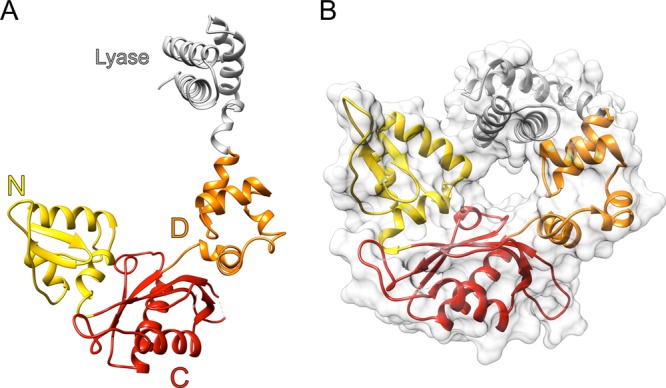
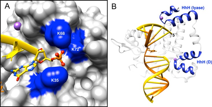
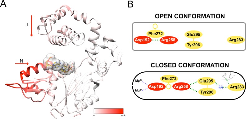
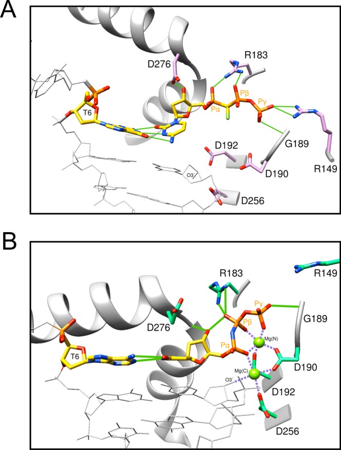
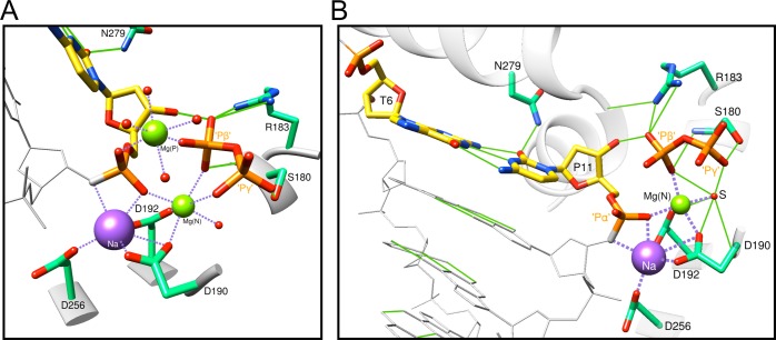
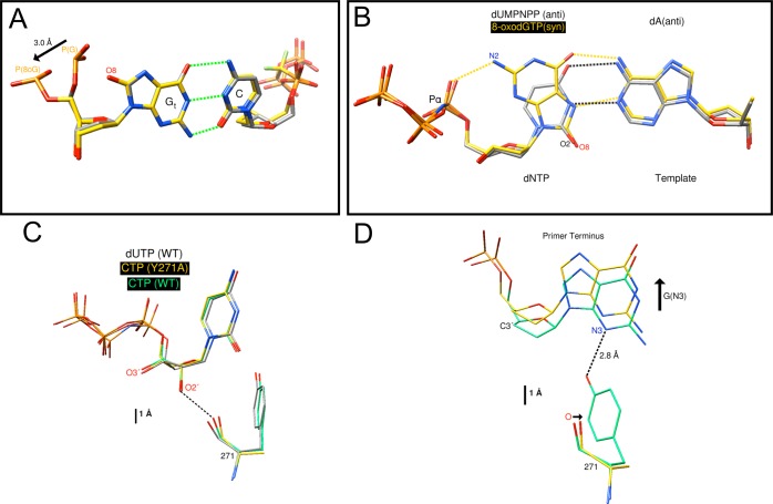
DNA polymerase (pol) β is a small eukaryotic DNA polymerase composed of two domains. Each domain contributes an enzymatic activity (DNA synthesis and deoxyribose phosphate lyase) during the repair of simple base lesions. These domains are termed the polymerase and lyase domains, respectively. Pol β has been an excellent model enzyme for studying the nucleotidyl transferase reaction and substrate discrimination at a molecular level. In this review, recent crystallographic studies of pol β in various liganded and conformational states during the insertion of right and wrong nucleotides as well as during the bypass of damaged DNA (apurinic sites and 8-oxoguanine) are described. Structures of these catalytic intermediates provide unexpected insights into mechanisms by which DNA polymerases enhance genome stability. These structures also provide an improved framework that permits computational studies to facilitate the interpretation of detailed kinetic analyses of this model enzyme.


Figures

Figure 1
Figure 1
Base excision repair. Base excision repair involves the removal of a damaged nucleotide (red pointers) from DNA. It is replaced with an undamaged nucleotide (green pointers). The damaged base is removed by a damage specific DNA glycosylase (1) that hydrolyzes the N-glycosidic bond between the deoxyribose and damaged base. In this image, uracil would be removed by uracil DNA glycosylase. AP endonuclease 1 incises the sugar–phosphate backbone 5′ to the AP site (2). The lyase domain of pol β (3) removes the 5′-dRP group (red pointer), and the polymerase domain inserts a nucleotide in a template-dependent reaction (green pointer). DNA ligase (4) seals the nicked DNA, resulting in restoration of the original DNA structure.
Scheme 1
Scheme 1. Minimal DNA Polymerase Reaction Pathway
After binding DNA (step 1), the nucleotide triphosphate binds, forming an initial ternary complex (circle, step 2). The polymerase–substrate complex undergoes rapid conformational adjustments that lead to a productive ternary substrate complex (square, step 3). Catalysis (step 4) leads to a postchemistry product ternary complex. Product release occurs concurrent with or following conformational changes (step 5) that facilitate PPi release (step 6). Translocation of the nascent base pair upstream vacating the active site prepares the polymerase for the next insertion event (gray solid line). Alternatively, product DNA+1 may dissociate from the polymerase (step 7), terminating DNA synthesis. While two divalent metals (Mg2+) are required for catalysis, they are not explicitly shown in this scheme. See the text for the role of these metals.
Figure 2
Figure 2
Domain and subdomain organization of DNA polymerase β. Ribbon representation of pol β illustrating the polymerase (colored) and amino-terminal lyase (gray) domains. The polymerase domain is composed of three subdomains: D, orange; C, red; and N, yellow. These correspond to the thumb, palm, and fingers subdomains, respectively, of DNA polymerases that utilize an architectural analogy to a right hand. (A) The structure of the apoenzyme indicates that it forms an extended structure (PDB entry 1BPD). (B) The molecular surface (semitransparent) of pol β bound to single-nucleotide gapped DNA (PDB entry 3ISB) exhibits a global doughnutlike structure in which the lyase domain interacts with the N-subdomain of the polymerase domain.
Figure 3
Figure 3
DNA binding. (A) Surface representation of the lyase domain highlighting the 5′-phosphate binding pocket (PDB entry 3ISB). The 5′-phosphate is hydrogen bonded to Lys35 (K35) and Lys68 (K68). Lys72 serves as the nucleophile for removal of the 5′-dRP intermediate during BER. (B) The lyase domain and the D-subdomain each have a HhH motif (blue ribbons) that interacts with the DNA backbone of the incised DNA strand (yellow) downstream and upstream of the gap, respectively. This motif binds a monovalent cation (Na+, purple) making sequence nonspecific interactions with the DNA backbone. The template strand is colored orange, and most of the nucleotides are illustrated in a ladder representation. The coding templating base in the gap is shown as sticks. The 5′-margin in the gap of the incised strand is indicated.
Figure 4
Figure 4
Nucleotide-induced conformational changes. (A) Ribbon representation of the single-nucleotide gapped DNA binary pol β complex (PDB entry 3ISB). The ribbon is colored according to the protein backbone displacement upon formation of the ternary substrate complex (PDB entry 2FMS), from white (0 Å) to red (10 Å). The nascent base pair is also illustrated with a semitransparent surface representation (PDB entry 2FMS). Significant domain and subdomain repositioning occurs exclusively in the lyase domain and N-subdomain. (B) In the open conformation, Arg283 (N-subdomain, yellow) does not interact with other key residues, but in the closed conformation, it interacts with the templating (coding) base, the upstream template nucleotide, and Glu295 (green dotted lines). Consequently, the position of the N-subdomain is structurally transmitted to the active site through a series of interactions involving Arg283 and Asp192 that coordinates (purple dotted lines) both active site Mg2+ ions. This is also accompanied by altered interactions of Glu295/Tyr296 with Arg258 in the open and closed forms. Phe272 is postulated to transiently interfere with interactions between Asp192 and Arg258, permitting an interaction with Glu295/Tyr296. Residues in the C-subdomain are colored red. Panel B was adapted from ref (40).
Figure 5
Figure 5
Nucleotide binding to pol β. (A) A ternary substrate complex with the correct incoming nucleotide was trapped in the open conformation using a mutant of pol β that destabilizes the closed conformation. In this structure (PDB entry 4F5N), protein side chains coordinate the extended anionic triphosphate moiety of the incoming nonhydrolyzable dCTP analogue, dCMP(CF2)PP. Although the nascent base pair (yellow carbon atoms) is severely buckled, the incoming cytosine base hydrogen bonds (green lines) with the templating guanine. Asp276 (D276) hydrogen bonds with O3′ of the incoming nucleotide, while Arg183 (R183) coordinates nonbridging oxygens on the α-phosphate (Pα) and β-phosphate (Pβ) of the incoming nucleotide. Arg149 (R149) and Gly189 (D189) coordinate the γ-phosphate (Pγ) of the incoming nucleotide. Active site aspartates (D190, D192, and D256) that coordinate active site metals are also indicated. The templating (coding) nucleotide and primer terminus are also indicated (T6 and O3′, respectively). (B) The active site structure and metal coordination of the precatalytic ternary substrate complex (PDB entry 2FMS) are consistent with a two-metal mechanism for nucleotidyl transfer. This closed structure was trapped with an inert dUTP analogue (dUMPNPP). Importantly, the primer terminus O3′ coordinates the catalytic Mg2+, labeled Mg(C). The catalytic Mg2+ also coordinates all three active site aspartates (purple dashed lines). In this structure, O3′ of the primer terminus is 3.4 Å from the α-phosphate of dUMPNPP. A nucleotide binding metal, Mg(N), coordinates nonbridging oxygens on all three phosphates. The protein coordination of the triphosphate also differs from that observed in the absence of metals. Arg183 now only coordinates the β-phosphate, and Arg149 has lost its direct contact with Pγ.
Figure 6
Figure 6
Structures of the closed product complex of pol β. (A) As the nucleotidyl transfer reaction proceeds in the crystal, an additional divalent metal is observed in the closed product complex. After a 40 s reaction in the crystal (PDB entry 4KLG), the active site structure reveals a new metal [Mg(P)] that bridges the two products, i.e., coordinates nonbridging oxygens on the phosphates of the incorporated dCMP and the remnant β-phosphate of PPi (yellow carbons). Water molecules (red spheres) complete the octahedral coordination (purple dashed lines). A sodium ion (purple sphere, Na+) replaces the catalytic magnesium, but the nucleotide-associated Mg2+ still coordinates nonbridging oxygens on the phosphates of the products (dCMP and PPi). (B) After an extended reaction in the crystal (45 min, PDB entry 4KLL), the polymerase remains closed, but the PPi appears to be preparing to dissociate. In this case, a water molecule (S) replaces the remnant of the γ-phosphate for nucleotide metal coordination. Hydrogen bonds are displayed as green lines.
Figure 7
Figure 7
Intermediate pol β structures for insertion of the wrong nucleotide. (A) Overlay of the ternary substrate complex structure with a correct incoming nucleotide (PDB entry 2FMS, light green carbons) with a precatalytic complex with an active site mismatch (PDB entry 3C2M, yellow carbons; dG-dAMPCPP)., The position of α-helix N (ribbon) of the N-subdomain indicates that the polymerase is in the closed conformation. The coding template base (T6) is shifted upstream 3.2 Å, while the incoming nucleotide is positioned in the dNTP binding pocket. The α-, β-, and γ-phosphates of the incoming nucleotide are denoted Pα, Pβ, and Pγ, respectively. The primer terminus (P10) of the mismatched structure rotates to follow its templating base that has shifted upstream, as the coding templating nucleotide vacates its binding site. This displaces O3′ from the primer terminus (highlighted), thereby deterring incorrect nucleotide insertion. (B) In contrast to insertion of the correct nucleotide in the crystal, misinsertion of an incorrect nucleotide results in an open binary complex in which PPi has dissociated (PDB entry 4KLU). The structure of the binary product complex following misinsertion indicates that the enzyme is in the open conformation (pink carbons). In this open conformation, the density for the misinserted nucleotide is poor, indicating that it can assume multiple conformations.
Figure 8
Figure 8
Structures of pol β with nonstandard substrates. (A) Structure of a ternary substrate complex with 8-oxoG in the templating position (Gt) paired with cytosine (PDB entry 3RJI, yellow carbons). It is superimposed with a ternary substrate complex with a templating guanine (PDB entry 2FMP, gray carbons). The bases are in an anti conformation, but the phosphate of the adducted guanine is repositioned to relieve steric and electrostatic clashes. (B) Structure of a ternary substrate complex with an incoming 8-oxodGTP in the syn conformation paired with adenine (PDB entry 3MBY, yellow carbons). In contrast to a staggered arrangement of bases described previously with active site mismatches, the 8-oxodGTP-dA mispair is planar because 8-oxodGTP assumes the syn conformation while the templating deoxyadenine (dA) remains in the anti conformation providing for good Watson–Crick geometry. The syn conformation is stabilized through Hoogsteen hydrogen bonding with the templating adenine (yellow dashed lines) and a hydrogen bond with Asn279 (not shown). The syn conformation of 8-oxodGTP positions O8 in the DNA minor groove in a position similar to that of O2 for a Watson–Crick base pair. The structure of the ternary substrate complex with dUMPNPP paired with adenine is shown for reference (PDB entry 2FMS, gray carbons). Additionally, an intramolecular hydrogen bond between N2 and a nonbridging oxygen on Pα (pro-SP) of 8-oxodGTP could stabilize the syn conformer. (C) Structures of precatalytic ternary substrate complexes of pol β with an incoming CTP (wild-type enzyme, PDB entry 3RH4, light green carbons; Y271A mutant, PDB entry 3RH6, yellow carbons) were superimposed with the wild-type enzyme with an incoming dUMPNPP (PDB entry 2FMS, gray carbons)., The 2′-ribose oxygen is unusually close to the carbonyl oxygen of Tyr271 (2.54 Å, dashed line) but is well-accommodated in the closed complex of these structures. Replacing the tyrosine side chain with a methyl group (Y271A) provides additional freedom to subtly displace this carbonyl from O2′, resulting in a mutant enzyme that displays a decreased level of discrimination for ribonucleotides. (D) Tyr271 hydrogen bonds to the base of the minor groove edge of the primer terminus (dashed black line). Substitution of Tyr271 with alanine results in the loss of this hydrogen bond and displacement of the dideoxy-terminated primer into the major groove. This is illustrated by examining the position of N3 of the guanine base in the structures of wild-type and mutant enzymes with an incoming CTP. The carbonyl of Ala271 is also illustrated.

References

    1. Braithwaite D. K.; Ito J. (1993) Compilation, alignment, and phylogenetic relationships of DNA polymerases. Nucleic Acids Res. 21, 787–802. - PMC - PubMed
    1. Beard W. A.; Wilson S. H. (2000) Structural design of a eukaryotic DNA repair polymerase: DNA polymerase β. Mutat. Res. 460, 231–244. - PubMed
    1. Beard W. A.; Wilson S. H. (2006) Structure and mechanism of DNA polymerase β. Chem. Rev. 106, 361–382. - PubMed
    1. Nakamura J.; Swenberg J. A. (1999) Endogenous apurinic/apyrimidinic sites in genomic DNA of mammalian tissues. Cancer Res. 59, 2522–2526. - PubMed
    1. Lindahl T.; Nyberg B. (1972) Rate of depurination of native deoxyribonucleic acid. Biochemistry 11, 3610–3618. - PubMed

Publication types