. 2014;11(4):334-8.
doi: 10.4161/rna.28372. Epub 2014 Mar 6.

The HIV-1 Tat protein modulates CD4 expression in human T cells through the induction of miR-222

Affiliations

The HIV-1 Tat protein modulates CD4 expression in human T cells through the induction of miR-222

Elisa Orecchini et al. RNA Biol. 2014.

Abstract

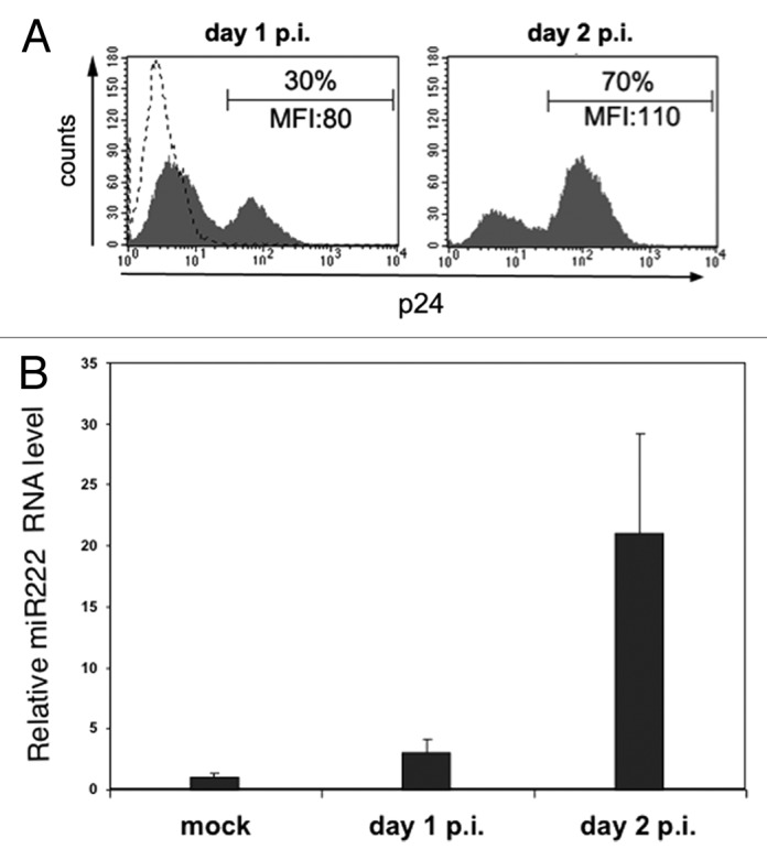
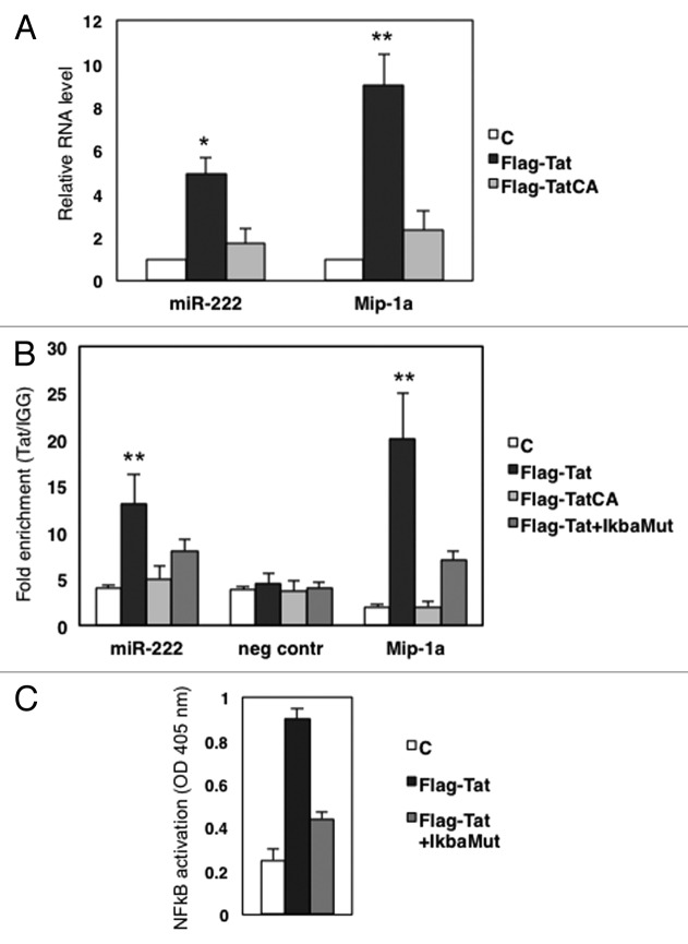
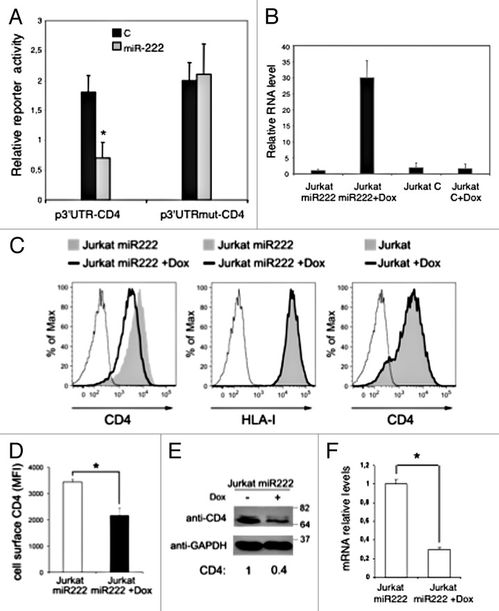
Several cellular microRNAs show substantial changes in expression during HIV-1 infection and their active role in the viral life cycle is progressively emerging. In the present study, we found that HIV-1 infection of Jurkat T cells significantly induces the expression of miR-222. We show that this induction depends on HIV-1 Tat protein, which is able to increase the transcriptional activity of NFkB on miR-222 promoter. Moreover, we demonstrate that miR-222 directly targets CD4, a key receptor for HIV-1, thus reducing its expression. We propose that Tat, by inducing miR-222 expression, complements the CD4 downregulation activity exerted by other viral proteins (i.e., Nef, Vpu, and Env), and we suggest that this represents a novel mechanism through which HIV-1 efficiently represses CD4 expression in infected cells.

Keywords: CD4; HIV-1; NFkB; microRNAs; post-transcriptional regulation.


Figures

None
Figure 1. HIV-1 infection induces miR-222 expression. Jurkat cells were infected with HIV-1 by standard spin-infection method or not infected (mock) and analyzed at day 1 and 2 post-infection (p.i.) by flow cytometry (A) and qRT-PCR (B). (A) Histograms show the fluorescence of cells labeled with PE-conjugated anti-p24 mAb. The mean fluorescence intensity (MFI) values and the percentage of p24+ cells are indicated. Dashed line represents labeled mock-infected cells. (B) qRT-PCR was used to measure endogenous miR-222 expression level. Relative miR-222 level in mock Jurkat cells was set to 1. Error bars are SD of one experiment performed in triplicate. One representative experiment out of three is shown.
None
Figure 2. Tat increases miR-222 levels in a NFkB-dependent manner. (A) HeLa cells were transfected with the empty vector (C), Flag-Tat or Flag-Tat CA expression constructs. Forty-eight hours post-transfection, total RNA was collected, and expression of miR-222 was analyzed by qRT-PCR. As a control, we checked the expression of MIP-1a, a NFkB-dependent gene that was shown to be sensitive to Tat. Results shown represent the average of three independent experiments, all performed in triplicate. (B) ChIP assay of chromatin isolated from HeLa cells transfected with empty plasmid (C), Flag Tat, Flag-TatCA, or Flag-Tat plus pCMV-IkBam plasmids (Flag-Tat+IkBa), and immunoprecipitated by anti-Flag or control IgGs, followed by qPCR analysis with primers targeted either to miR-222 enhancer region, or to a sequence on chromosome 1 used as the negative control, or to Mip-1a promoter used as positive control. The data show occupancy relative to control IgGs and represent mean ± SD of three independent experiments. (C) Nuclear protein extracts from HeLa cells transfected with empty plasmid (C), Flag Tat, Flag-Tat plus pCMV-IkBam plasmids (Flag-Tat+IkBa), were harvested and p65 NFκB activity was measured by transAM p65 protein kit. *, P < 0.05; **, P < 0.01 by Student's t test
None
Figure 3. CD4 mRNA is a target of miR-222. (A) p3UTR-CD4 or p3UTRmut-CD4 luciferase constructs containing a wild-type (black columns) or a mutated (gray columns) CD4 3UTR were co-transfected into HeLa cells with empty vector (C), or plasmid expressing miR-222 (p-222). Luciferase activity was determined 48 h after transfection. The ratio of normalized sensor to control luciferase activity is shown. Error bars represent SD and were obtained from three independent experiments. (B) Jurkat miR-222 and Jurkat C cells treated or not with DOX for 48 h, were analyzed by qRT-PCR to measure miR-222 expression level. Relative miR-222 level in Jurkat miR-222 cells not induced by DOX was set to 1. Error bars are SD of one experiment performed in triplicate. (C–F) Jurkat miR-222 cells treated as described in (B) were analyzed by flow cytometry (C and D), western blotting (E), and qRT-PCR (F). (C) Histograms show the fluorescence distribution of cells labeled with PE-conjugated anti-CD4 (left and right panels) or anti-HLA-I (middle panel) mAbs. Also, parental Jurkat cells with and without DOX were analyzed for cell-surface CD4 expression (right panel). Solid lines and filled gray histograms represent cells with and without DOX, respectively. Staining with control IgG is also shown (thin line). (D) The cell-surface CD4 MFI ± SEM obtained in three independent experiments like the one in panel (A) is shown. (E) Equal amounts of total cell lysates (20 μg) were separated by 10% SDS-PAGE followed by immunoblotting with anti-CD4 and anti-GAPDH antibodies. Molecular weight standards are indicated (kDa). The CD4 levels were measured by densitometry, normalized by GAPDH, and expressed by setting at 1 the value found in untreated Jurkat miR-222 cells. (F) qRT-PCR was used to assess CD4 mRNA expression. Relative mRNA level in untreated Jurkat miR-222 cells was set at 1. Error bars are SD of an experiment performed in triplicate. Results shown in panels (C–F) are representative of three independent experiments. *P < 0.05 by Student’s t test.

References

    1. Bartel DP. MicroRNAs: target recognition and regulatory functions. Cell. 2009;136:215–33. doi: 10.1016/j.cell.2009.01.002. - DOI - PMC - PubMed
    1. Swaminathan S, Murray DD, Kelleher AD. miRNAs and HIV: unforeseen determinants of host-pathogen interaction. Immunol Rev. 2013;254:265–80. doi: 10.1111/imr.12077. - DOI - PubMed
    1. Huang J, Wang F, Argyris E, Chen K, Liang Z, Tian H, Huang W, Squires K, Verlinghieri G, Zhang H. Cellular microRNAs contribute to HIV-1 latency in resting primary CD4+ T lymphocytes. Nat Med. 2007;13:1241–7. doi: 10.1038/nm1639. - DOI - PubMed
    1. Nathans R, Chu CY, Serquina AK, Lu CC, Cao H, Rana TM. Cellular microRNA and P bodies modulate host-HIV-1 interactions. Mol Cell. 2009;34:696–709. doi: 10.1016/j.molcel.2009.06.003. - DOI - PMC - PubMed
    1. Triboulet R, Mari B, Lin YL, Chable-Bessia C, Bennasser Y, Lebrigand K, Cardinaud B, Maurin T, Barbry P, Baillat V, et al. Suppression of microRNA-silencing pathway by HIV-1 during virus replication. Science. 2007;315:1579–82. doi: 10.1126/science.1136319. - DOI - PubMed

Publication types