. 2014 Apr 24;508(7497):526-30.
doi: 10.1038/nature13242. Epub 2014 Apr 9.

Trogocytosis by Entamoeba histolytica contributes to cell killing and tissue invasion

Affiliations

Trogocytosis by Entamoeba histolytica contributes to cell killing and tissue invasion

Katherine S Ralston et al. Nature. .

Abstract

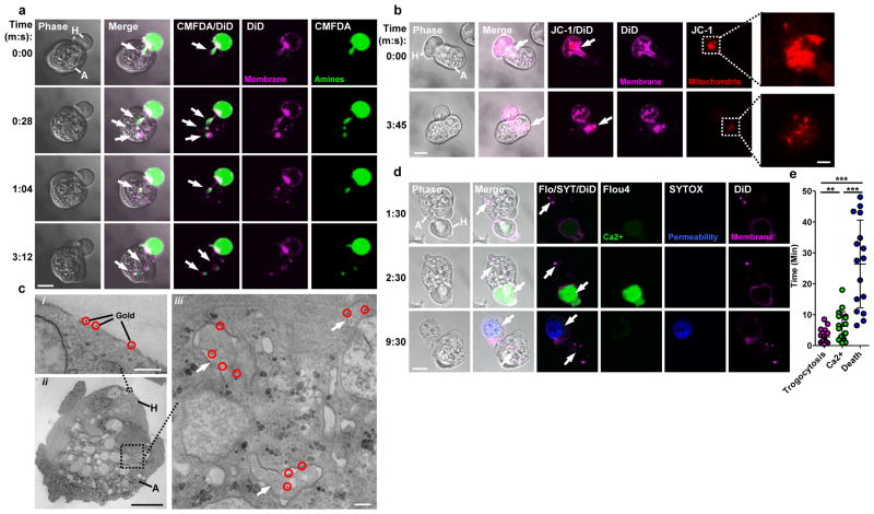
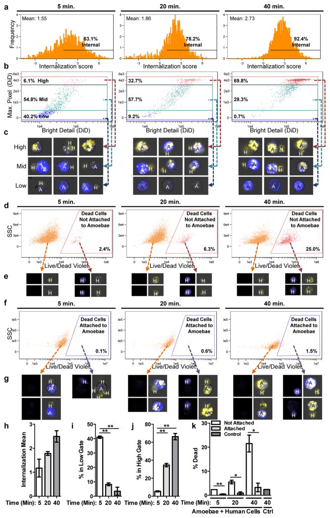
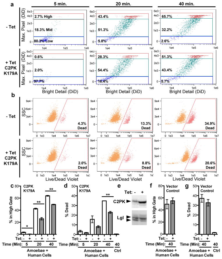
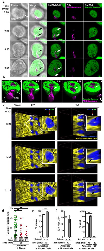
Entamoeba histolytica is the causative agent of amoebiasis, a potentially fatal diarrhoeal disease in the developing world. The parasite was named "histolytica" for its ability to destroy host tissues, which is probably driven by direct killing of human cells. The mechanism of human cell killing has been unclear, although the accepted model was that the parasites use secreted toxic effectors to kill cells before ingestion. Here we report the discovery that amoebae kill by ingesting distinct pieces of living human cells, resulting in intracellular calcium elevation and eventual cell death. After cell killing, amoebae detach and cease ingestion. Ingestion of human cell fragments is required for cell killing, and also contributes to invasion of intestinal tissue. The internalization of fragments of living human cells is reminiscent of trogocytosis (from Greek trogo, nibble) observed between immune cells, but amoebic trogocytosis differs because it results in death. The ingestion of live cell material and the rejection of corpses illuminate a stark contrast to the established model of dead cell clearance in multicellular organisms. These findings change the model for tissue destruction in amoebiasis and suggest an ancient origin of trogocytosis as a form of intercellular exchange.


Conflict of interest statement

The authors declare that no competing financial interests exist.

Figures

Figure 1
Figure 1. Amoebae internalize human cell bites, preceding human cell death
a, DiD and CMFDA-labeled human Jurkat cells (H); an amoeba (A) internalizes bites (arrows) over time. Images are representative of three independent experiments. b, DiD and JC-1-labeled human Jurkat cells; mitochondria (arrow) are ingested by the amoeba in a bite. Images are representative of six independent experiments. c, i–ii, Electron microscopy with gold-labeled human Jurkat cells (gold, circles). iii, Bites (arrows) within an amoeba. Images are representative of two independent experiments. d, DiD and Flou4-labeled human Jurkat cells; with SYTOX blue present during imaging. Arrows, amoebic trogocytosis (1:30), [Ca2+]i elevation (2:30) and membrane permeabilization (9:30). Images are representative of 15 independent experiments. e, Timing in 60 cells from 15 independent experiments; shown are data points, means and standard deviations; P-values from t-tests: * < .05, ** < .01, *** < .001. Bars: 10 μm (a, b, d), 2 μm (b, insets), 0.5 μm (c, i, iii) and 5 μm (c, ii).
Figure 2
Figure 2. Amoebic trogocytosis is predominant and specific to live human cells
Imaging flow cytometry with CMFDA-labeled amoebae (A, blue), DiD-labeled human Jurkat cells (H, yellow) and Live/Dead Violet-labeled dead cells (purple). a, Detection of internalized human material. b, Measurement of fragmentation of internalized human material, gated from low to high. c, Example merged CMFDA, DiD and bright field images. Images are representative of 10,000 images collected for each replicate. d, Detection of dead cells not attached to amoebae. e, Example images; Live/Dead Violet (left panels) and merged CMFDA, DiD and bright field (right panels). Images are representative of 10,000 images collected for each replicate. f, Detection of dead cells attached to amoebae. g, Example images, as in e. Images are representative of 10,000 images collected for each replicate. h – k Means and standard deviations for biological replicates (10,000 events/each). P-values from t-tests: * < .05, ** < .01, *** < .001.
Figure 3
Figure 3. Amoebic trogocytosis contributes to human cell killing
a – d, Imaging flow cytometry analysis with amoebae expressing a tetracycline (tet)-inducible copy of EhC2PK K179A. a, Measurement of fragmentation. b, Detection of dead cells. c – d, Means and standard deviations for biological duplicates at each time point (10,000 events/each). e, Western blot demonstrating EhC2PK over-expression in tet-induced cells; Lgl, loading control. Blots are representative of three independent experiments. f – g, Imaging flow cytometry analysis with vector control amoebae; shown are means and standard deviations for biological duplicates (10,000 events/each). P-values from t-tests: * < .05, ** < .01, *** < .001.
Figure 4
Figure 4. Amoebic trogocytosis occurs with red blood cells, contributes to intestinal invasion, and promotes enhanced cell killing
a, DiD-labeled red blood cells (RBC) and CMFDA-labeled amoebae (A); ingested bites (arrows) and a fragment remains extracellular (0:51). Bar, 5 μm. Images are representative of three independent experiments. b, Example 3-D reconstruction with DiD-labeled red blood cells and CMFDA-labeled amoebae; biting (arrows) and a fragment remains extracellular (3:25). Images are representative of three independent experiments. c, Planes from 4-D two-photon microscopy with mouse intestinal tissue (yellow); calcein violet-labeled amoebae (blue) are ingesting bites (arrowheads). Images are representative of three independent experiments. d, Amoebae treated with wortmannin or cytochalasin D were incubated with tissue. Invasion depth, means and standard deviations from biological replicates (at least 40 cells/treatment); P-values are indicated. e–g, Amoebae were incubated with or without human Jurkat cells (“primed”), prior to incubation with labeled human Jurkat cells. e, Measurement of internalization. f, Measurement of fragmentation. g, Detection of dead cells. Means and standard deviations for biological duplicates (10,000 events/each). P-values from t-tests: * < .05, ** < .01, *** < .001.

Comment in

References

    1. Ralston KS, Petri WA., Jr Tissue destruction and invasion by Entamoeba histolytica. Trends Parasitol. 2011;27:254–263. - PMC - PubMed
    1. Batista FD, Iber D, Neuberger MS. B cells acquire antigen from target cells after synapse formation. Nature. 2001;411:489–494. - PubMed
    1. Huang JF, et al. TCR-Mediated internalization of peptide-MHC complexes acquired by T cells. Science. 1999;286:952–954. - PubMed
    1. Hudrisier D, Riond J, Mazarguil H, Gairin JE, Joly E. Cutting edge: CTLs rapidly capture membrane fragments from target cells in a TCR signaling-dependent manner. J Immunol. 2001;166:3645–3649. - PubMed
    1. Hudson L, Sprent J, Miller JF, Playfair JH. B cell-derived immunoglobulin on activated mouse T lymphocytes. Nature. 1974;251:60–62. - PubMed

Publication types