. 2014 May 29;509(7502):617-21.
doi: 10.1038/nature13250. Epub 2014 Apr 6.

Epidermal Merkel cells are mechanosensory cells that tune mammalian touch receptors

Affiliations

Epidermal Merkel cells are mechanosensory cells that tune mammalian touch receptors

Srdjan Maksimovic et al. Nature. .

Abstract

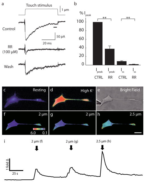
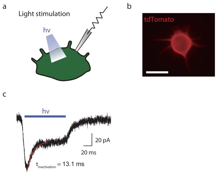
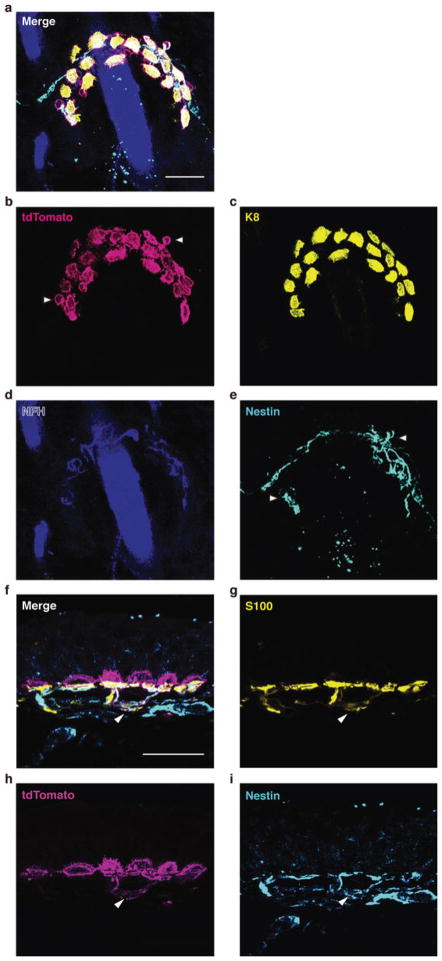
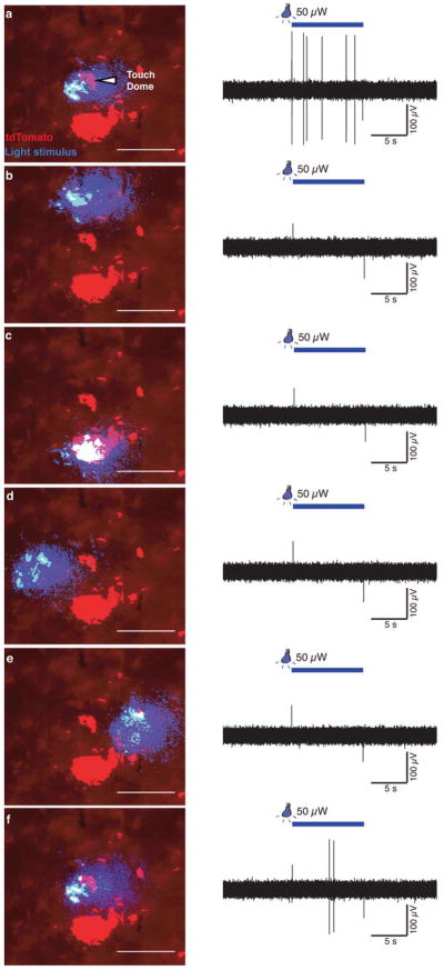
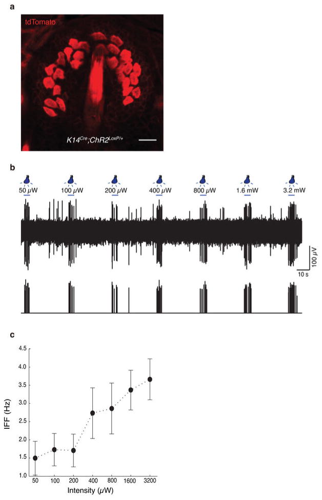
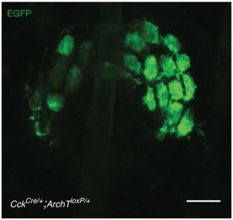
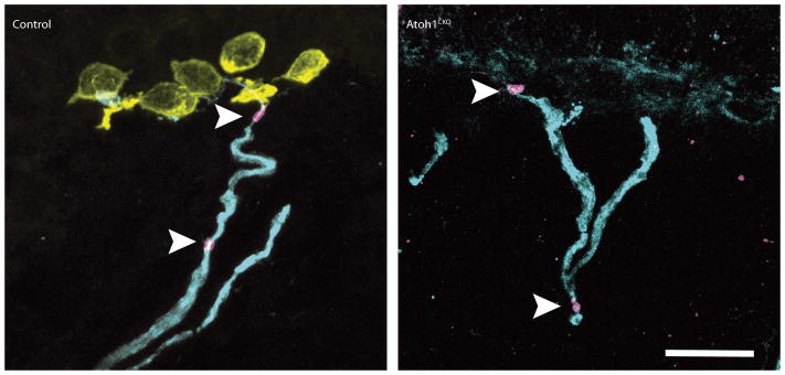
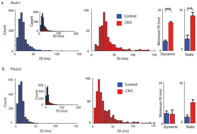
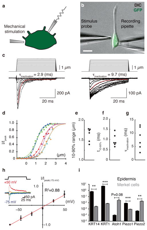
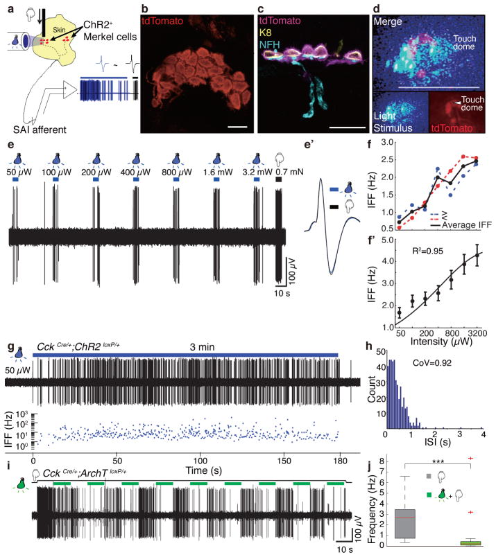
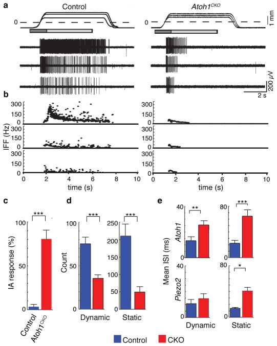
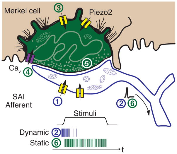
Touch submodalities, such as flutter and pressure, are mediated by somatosensory afferents whose terminal specializations extract tactile features and encode them as action potential trains with unique activity patterns. Whether non-neuronal cells tune touch receptors through active or passive mechanisms is debated. Terminal specializations are thought to function as passive mechanical filters analogous to the cochlea's basilar membrane, which deconstructs complex sounds into tones that are transduced by mechanosensory hair cells. The model that cutaneous specializations are merely passive has been recently challenged because epidermal cells express sensory ion channels and neurotransmitters; however, direct evidence that epidermal cells excite tactile afferents is lacking. Epidermal Merkel cells display features of sensory receptor cells and make 'synapse-like' contacts with slowly adapting type I (SAI) afferents. These complexes, which encode spatial features such as edges and texture, localize to skin regions with high tactile acuity, including whisker follicles, fingertips and touch domes. Here we show that Merkel cells actively participate in touch reception in mice. Merkel cells display fast, touch-evoked mechanotransduction currents. Optogenetic approaches in intact skin show that Merkel cells are both necessary and sufficient for sustained action-potential firing in tactile afferents. Recordings from touch-dome afferents lacking Merkel cells demonstrate that Merkel cells confer high-frequency responses to dynamic stimuli and enable sustained firing. These data are the first, to our knowledge, to directly demonstrate a functional, excitatory connection between epidermal cells and sensory neurons. Together, these findings indicate that Merkel cells actively tune mechanosensory responses to facilitate high spatio-temporal acuity. Moreover, our results indicate a division of labour in the Merkel cell-neurite complex: Merkel cells signal static stimuli, such as pressure, whereas sensory afferents transduce dynamic stimuli, such as moving gratings. Thus, the Merkel cell-neurite complex is an unique sensory structure composed of two different receptor cell types specialized for distinct elements of discriminative touch.


Figures

Extended Data Figure 1
Extended Data Figure 1. a–b, Mechanically activated currents in Merkel cells were inhibited by Ruthenium Red (RR)
a, Representative trace of mechanically evoked current induced by 1-μm mechanical displacement. Application of RR (100 μM) attenuated mechanically activated current. b, Peak currents (Ipeak) were estimated from 250 μs around peak and steady-state currents (Iss) were estimated from the last 5 ms (black bar in 1a) of mechanical displacements. Data were normalized by Ipeak for each cell. With Ruthenium Red, Ipeak was reduced to 38±7% of control condition. Steady state currents were also reduced by RR (N=4; control: 9±1% of Ipeak; RR: 2±1% of Ipeak). c–i, Merkel cells display reversible Ca2+ responses to focal displacements applied to somata. c, Representative pseudocolor images of fura-2 ratios (340:380) of a Merkel cell at rest. d, A Merkel cell activated by depolarizing (high-K+) solution. e, A brightfield image showing the position of the stimulus probe. f–h, Peak responses corresponding to each displacement. ‘Fold Δ’ is the fold change in fluorescence ratio from baseline. Scale bar, 10 μm. i, Representative time course of mean fura-2 ratios during the touch stimuli shown above. Stimulus onset in f–h is indicated by arrows. Calcium responses were stimulus dependent. Similar responses were observed from 11 Merkel cells.
Extended Data Figure 2
Extended Data Figure 2. ChR2+ Merkel cells display light-activated inward currents
a, Light-activated currents were recorded with whole-cell, tight-seal voltage clamp methods. b, Fluorescent image of a ChR2-tdTomato expressing Merkel cell. Scale bar, 10 μm. c, Representative trace for light-activated inward currents at a holding potential of −70 mV. Inactivation kinetics were measured by fitting a single exponential curve (red).
Extended Data Figure 3
Extended Data Figure 3. Immunostaining of ChR2-expressing touch domes
a–e, Whole-mount staining and confocal axial projection of the touch dome shown in Fig. 2d. a, Merged image. b–d, Expression of ChR2-tdTomato was present in Merkel cells (Keratin-8, K8), but absent from sensory terminals (Neurofilament Heavy, NFH). e, Some terminal Schwann cells (Nestin) also expressed ChR2 (arrowheads in b & e). f–i, Immunostaining of skin cryosections. f, Merged image. g–i, ChR2-tdTomato was present in some S100+ Schwann cells that also expressed Nestin, a marker for type II terminal Schwann cells (arrowheads in f–i). Scale bars, 20 μm.
Extended Data Figure 4
Extended Data Figure 4. Light-evoked activity is specific to touch-dome illumination
a & f, Responses to light stimuli centred on a touch dome. b–e, When the light stimulus was positioned around the touch dome, no light-evoked activity is observed. Illuminating a cluster of ChR2+ dermal cells did not evoke any responses (c). f, To confirm that the absence of light-evoked activity was not due to the loss of Merkel cells and/or neuronal fibres, the experiment ended by re-positioning the light stimulus over the touch dome to re-elicit light-evoked activity. Images have been thresholded for clarity. Scale bars, 200 μm.
Extended Data Figure 5
Extended Data Figure 5. K14Cre;ChR2loxP/+ mice exhibit light-evoked SAI activity
a, Confocal image of a touch dome illustrating ChR2-tdTomato expression driven by K14Cre. ChR2-tdTomato expressed much stronger in Merkel cells than in neighbouring keratinocytes. b, Light-evoked responses from the touch dome shown in a to seven light intensities as indicated. Spike sorting and clustering analysis were used to identify the unit that fired in phase with light (lower trace with spike positions and their amplitudes). c, Mean IFFs for light with varying illumination intensities on a log-intensity scale (N=3 recordings). Scale bar, 20 μm.
Extended Data Figure 6
Extended Data Figure 6. Confocal axial projection of a touch dome shows selective ArchT-EGFP expression in Merkel cells driven by CckCre
ArchT-EGFP expression was not observed in touch-dome afferents. Scale bar, 20 μm.
Extended Data Figure 7
Extended Data Figure 7. Structure of touch-dome afferents in Atoh1CKO mice
Immunostaining of skin cryosections from Atoh1CKO and control genotypes are shown. Antibodies labelling myelinated afferents (NFH; cyan), Merkel cells (Keratin-8; yellow), nodes of Ranvier (βIV spectrin; magenta) show that the general structure of touch-dome afferents, including myelinated branches and Nodes of Ranvier (arrowheads), appears normal even in the absence of Merkel cells. Cell nuclei are labelled with DAPI (blue). Scale bar, 20 μm.
Extended Data Figure 8
Extended Data Figure 8. Comparison of ISI distributions in Atoh1CKO, Piezo2CKO, and control genotypes
a, Histogram of ISI distribution during saturating responses in Atoh1CKO (Mean±SD, 43.4±59.2 ms, Median: 29.8 ms; n=466 intervals from N=6 units) and control genotypes (Mean±SD, 16.5±12.9 ms, Median: 13.8 ms; n=1412 intervals from N=5 units). Inset on the left illustrates all ISIs, including those >150 ms, which were excluded from the main histograms [14/466 intervals in Atoh1CKO and 1/1412 in control genotypes]. At right, bar graphs shows the minimum ISIs during dynamic and static phases. Minimum ISIs were longer in Atoh1CKO than control mice for both phases, indicating a loss of high-frequency firing during dynamic stimuli and static displacement (**P<0.02, ***P<0.01; Student’s t test). Mann-Whitney tests indicated that median values were also significantly different (P<0.001). b, Histogram of ISI distribution for Piezo2CKO (Mean±SD, 41.9 ±32.3 ms, Median: 23.4 ms; n=792 intervals from N=6 units) and control genotypes (Mean±SD, 13.9±1.4 ms, Median: 11.8 ms; n=1845 intervals from N=5 units). Main histograms excluded long intervals (>150 ms; 4/792 intervals in Piezo2CKO and 2/1845 in control mice.) Minimum ISIs were not significantly different in the dynamic phase (P≥0.76; Student’s t test and Mann-Whitney test); indicating that high-frequency firing is preserved in touch-dome afferents in these mice. For static phase firing, the means were not significantly different (P=0.095; Student’s t test); however non-parametric analysis indicated that medians differed between genotypes (P=0.0043; Mann Whitney test).
Figure 1
Figure 1. Merkel cells exhibit touch-evoked ionic currents and preferentially express Piezo2
a, Schematic of whole-cell recordings from dissociated Merkel cells. b, Merkel cells were identified based on GFP fluorescence and were stimulated by a glass probe driven by piezoelectric actuator. Scale bar, 10 μm. c, Representative traces of mechanosensitive currents from two Merkel cells (Vhold=−70 mV). Inactivation kinetics were estimated from single-exponential fits (red lines). d, Current-displacement relationships from individual Merkel cells (N=6; indicated by distinct colors). Currents were normalized to peak and fitted with Boltzmann functions (R2 > 0.97). e–g, Activation and inactivation kinetics and operating ranges for individual Merkel cells. h, Current-voltage relationship of mechanosensitive currents (N=5). Peak current at each holding potential was normalized to peak current at −75 mV. Reversal potential was estimated by linear regression (red line; R2 = 0.88). Inset: representative traces at each holding potential (denoted by colors). i, qPCR analysis of Piezo1 and Piezo2 transcripts in purified Merkel cells and epidermis. Markers of keratinocytes (KRT14 and KRT1) and Merkel cells (Atoh1) verified selective enrichment of Merkel cells. **P<0.0001, ***P≤0.001 (N=4).
Figure 2
Figure 2. Merkel cells are necessary and sufficient to elicit sustained action-potential trains in touch-dome afferents
a, Schematic of mouse ex vivo skin-nerve recordings. b, Confocal image of a ChR2-expressing touch dome in a living skin-nerve preparation. Scale bar, 20 μm. c, Immunostaining of skin cryosections shows expression of ChR2-tdTomato in Merkel cells (Keratin-8, K8) but not in touch-dome afferents (Neurofilament heavy, NFH). Scale bar, 20 μm. d, During electrophysiological recording, ChR2-expressing Merkel cells and blue-light stimuli were imaged separately using different filter sets (bottom insets). Merged image illustrates the illuminated area (top panel). Confocal reconstruction of this touch dome is in Extended Data Fig. 2a–e. Scale bar, 200 μm. e, Light pulses of increasing intensities elicited phase-locked action potentials from the touch dome in d. e, Comparison of spike shapes evoked by light (blue) and touch (black) confirmed single-unit recording. f, Mean instantaneous firing frequency (IFF) versus light intensity for a single touch-dome afferent. Blue trace shows mean IFFs from e. Red trace shows mean IFFs evoked by light intensities presented in decreasing order. The averages of these stimuli (black) were analysed further. f, Mean IFFs on a log-intensity scale (N=12 single units). Data were fit with a four-parameter Weibull sigmoidal function (R2=0.95). g–h, A sustained light-evoked response from the touch-dome afferent in d–e with corresponding ISI histogram. i–j, Optogenetic silencing of ArchT-expressing Merkel cells. i, Representative 3-min recording. j, Box-plot of firing rates during light-off (N=3 units, n=20 10-s periods) and light-on (same units, n=23 10-s periods). Two outliers are firing rates from initial light-on periods. P=0.001.
Figure 3
Figure 3. Atoh1CKO and Piezo2CKO mice show intermediately adapting (IA) responses
a, Mechanically evoked responses from touch-dome afferents in control and Atoh1CKO (K14Cre;Atoh1LacZ/flox) mice. Top trace shows ramp-and-hold displacements at three magnitudes with corresponding action potential trains below. Dashed line marks the point of skin contact (0 mm). Boxes indicate dynamic (dark gray: 1.5 s after displacement command onset) and static phases for analysis (light gray: 4 s after the beginning of hold command). b, IFFs of the responses in a. c, Proportion of IA responses to supra-threshold displacements in touch-dome afferents (N=5 control and N=6 Atoh1CKO units). d, Mean number of spikes in dynamic and static phases of saturating responses. e, Mean interspike intervals (ISIs) of Atoh1CKO (N=6 units), Piezo2CKO (N=6 units) and their respective control genotypes (N=5 units per genotype). Asterisks indicate statistically significant differences between mutant and control genotypes in Fisher’s exact test (c) and Student’s t test (d–e). *P<0.05; **P<0.02; ***P<0.01.
Figure 4
Figure 4. Model of active Merkel-cell inputs in touch reception
Deformation of the skin opens mechanotransduction channels in SAI afferents (1) to initiate action potential firing at the onset of dynamic stimuli (2). The presence of Merkel cells boosts dynamic firing through Piezo2-independent mechanisms. Skin deformation simultaneously activates Piezo2-dependent mechanotransduction channels in Merkel cells (3) to depolarize these cells, which produces calcium entry (4) and release of unidentified neurotransmitters (5) that trigger sustained firing (6). Schematic after Iggo and Muir.

Comment in

References

    1. Johnson KO. The roles and functions of cutaneous mechanoreceptors. Curr Opin Neurobiol. 2001;11:455–461. - PubMed
    1. Lumpkin EA, Caterina MJ. Mechanisms of sensory transduction in the skin. Nature. 2007;445:858–865. - PubMed
    1. Kwan KY, Glazer JM, Corey DP, Rice FL, Stucky CL. TRPA1 modulates mechanotransduction in cutaneous sensory neurons. The Journal of neuroscience. 2009;29:4808–4819. - PMC - PubMed
    1. Haeberle H, et al. Molecular profiling reveals synaptic release machinery in Merkel cells. Proc Natl Acad Sci USA. 2004;101:14503–14508. - PMC - PubMed
    1. Maksimovic S, Baba Y, Lumpkin EA. Neurotransmitters and synaptic components in the Merkel cell-neurite complex, a gentle-touch receptor. Ann NY Acad Sci. 2013;1279:13–21. - PMC - PubMed

Publication types

Substances